 Movie
Movie Controller

[English] 日本語
 Yorodumi
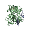
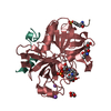
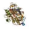
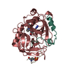
- PDB-1k5p: Hydrolytic haloalkane dehalogenase LINB from sphingomonas paucimo... -
+ Open data

- Basic information
| Entry | Database: PDB / ID: 1k5p | ||||||
|---|---|---|---|---|---|---|---|
| Title | Hydrolytic haloalkane dehalogenase LINB from sphingomonas paucimobilis UT26 at 1.8A resolution | ||||||
 Components Components | 1,3,4,6-tetrachloro-1,4-cyclohexadiene hydrolase | ||||||
 Keywords Keywords | HYDROLASE / DEHALOGENASE / LINDANE / BIODEGRADATION / ALPHA/BETA-HYDROLASE | ||||||
| Function / homology |  Function and homology information Function and homology informationhaloalkane dehalogenase / haloalkane dehalogenase activity / response to toxic substance / periplasmic space Similarity search - Function | ||||||
| Biological species |  Sphingomonas paucimobilis (bacteria) Sphingomonas paucimobilis (bacteria) | ||||||
| Method |  X-RAY DIFFRACTION / MOLECULAR REPLACEMENT / Resolution: 1.8 Å X-RAY DIFFRACTION / MOLECULAR REPLACEMENT / Resolution: 1.8 Å | ||||||
 Authors Authors | Streltsov, V.A. / Damborsky, J. / Wilce, M.C.J. | ||||||
 Citation Citation | Journal: Biochemistry / Year: 2003 Title: Haloalkane dehalogenase LinB from Sphingomonas paucimobilis UT26: X-ray crystallographic studies of dehalogenation of brominated substrates Authors: Streltsov, V.A. / Prokop, Z. / Damborsky, J. / Nagata, Y. / Oakley, A. / Wilce, M.C.J. | ||||||
| History |
|
- Structure visualization
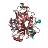
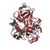
| Structure viewer | Molecule: MolmilJmol/JSmol |
|---|
- Downloads & links
-Download
| PDBx/mmCIF format | 1k5p.cif.gz | 83 KB | Display | PDBx/mmCIF format |
|---|---|---|---|---|
| PDB format | pdb1k5p.ent.gz | 60 KB | Display | PDB format |
| PDBx/mmJSON format | 1k5p.json.gz | Tree view | PDBx/mmJSON format | |
| Others |  Other downloads Other downloads |
-Validation report
| Arichive directory | https://data.pdbj.org/pub/pdb/validation_reports/k5/1k5pftp://data.pdbj.org/pub/pdb/validation_reports/k5/1k5p | HTTPS FTP |
|---|
-Related structure data
| Related structure data | 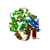 1iz7C 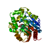 1iz8C 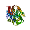 1k63C 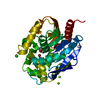 1k6eC  1cv2S S: Starting model for refinement C: citing same article ( |
|---|---|
| Similar structure data |


-Links
 PDBj
PDBj


- Assembly
| Deposited unit | 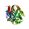
| ||||||||
|---|---|---|---|---|---|---|---|---|---|
| 1 |
| ||||||||
| Unit cell |
|
-Components
| #1: Protein | Mass: 33013.410 Da / Num. of mol.: 1 Source method: isolated from a genetically manipulated source Source: (gene. exp.) Sphingomonas paucimobilis (bacteria) / Plasmid: PMYLB1 / Production host: References: UniProt: P51698, UniProt: D4Z2G1*PLUS, haloalkane dehalogenase | ||||
|---|---|---|---|---|---|
| #2: Chemical |   Mass: 35.453 Da / Num. of mol.: 2 / Source method: obtained synthetically / Formula: Cl Mass: 35.453 Da / Num. of mol.: 2 / Source method: obtained synthetically / Formula: Cl#3: Chemical |   Mass: 24.305 Da / Num. of mol.: 3 / Source method: obtained synthetically / Formula: Mg Mass: 24.305 Da / Num. of mol.: 3 / Source method: obtained synthetically / Formula: Mg#4: Water | ChemComp-HOH / |  Mass: 18.015 Da / Num. of mol.: 423 / Source method: isolated from a natural source / Formula: H2O Mass: 18.015 Da / Num. of mol.: 423 / Source method: isolated from a natural source / Formula: H2O |
-Experimental details
-Experiment
| Experiment | Method: X-RAY DIFFRACTION / Number of used crystals: 1 |
|---|
- Sample preparation
| Crystal | Density Matthews: 1.73 Å3/Da / Density % sol: 28.23 % |
|---|---|
| Crystal grow | Temperature: 298 K / Method: vapor diffusion, sitting drop Details: PEG 4000, TRIS, magnesium chloride, VAPOR DIFFUSION, SITTING DROP, temperature 298K |
-Data collection
| Diffraction | Mean temperature: 100 K |
|---|---|
| Diffraction source | Source: ROTATING ANODE / Type: RIGAKU RU200 / Wavelength: 1.5418 Å |
| Detector | Type: MARRESEARCH / Detector: IMAGE PLATE / Date: Dec 14, 2000 / Details: mirrors |
| Radiation | Monochromator: Ni FILTER / Protocol: SINGLE WAVELENGTH / Monochromatic (M) / Laue (L): M / Scattering type: x-ray |
| Radiation wavelength | Wavelength: 1.5418 Å / Relative weight: 1 |
| Reflection | Resolution: 1.8→34.77 Å / Num. all: 21294 / Num. obs: 21294 / % possible obs: 83.7 % / Observed criterion σ(F): 0 / Observed criterion σ(I): 0 / Redundancy: 12 % / Biso Wilson estimate: 14.1 Å2 / Rmerge(I) obs: 0.077 / Rsym value: 0.077 / Net I/σ(I): 10.6 |
| Reflection shell | Resolution: 1.8→1.98 Å / Redundancy: 2.3 % / Rmerge(I) obs: 0.28 / Mean I/σ(I) obs: 2.2 / Num. unique all: 3393 / Rsym value: 0.28 / % possible all: 60.3 |
- Processing
| Software |
| |||||||||||||||||||||||||
|---|---|---|---|---|---|---|---|---|---|---|---|---|---|---|---|---|---|---|---|---|---|---|---|---|---|---|
| Refinement | Method to determine structure: MOLECULAR REPLACEMENT Starting model: PDB ENTRY 1CV2 Resolution: 1.8→34.77 Å / Rfactor Rfree error: 0.005 / Data cutoff high absF: 325933.63 / Data cutoff low absF: 0 / Isotropic thermal model: RESTRAINED / Cross valid method: THROUGHOUT / σ(F): 0 / σ(I): 0 / Details: The structure was refined also with XTALVIEW.
| |||||||||||||||||||||||||
| Solvent computation | Solvent model: FLAT MODEL / Bsol: 102.629 Å2 / ksol: 0.409013 e/Å3 | |||||||||||||||||||||||||
| Displacement parameters | Biso mean: 15.9 Å2
| |||||||||||||||||||||||||
| Refine analyze |
| |||||||||||||||||||||||||
| Refinement step | Cycle: LAST / Resolution: 1.8→34.77 Å
| |||||||||||||||||||||||||
| Refine LS restraints |
| |||||||||||||||||||||||||
| LS refinement shell | Resolution: 1.8→1.98 Å / Rfactor Rfree error: 0.017 / Total num. of bins used: 4
| |||||||||||||||||||||||||
| Xplor file |
|
