 Movie
Movie Controller


+ Open data

- Basic information
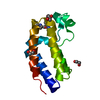
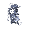
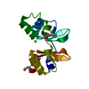
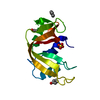
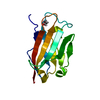
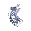
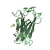
| Entry | Database: PDB / ID: 1k5a | ||||||
|---|---|---|---|---|---|---|---|
| Title | Crystal structure of human angiogenin double variant I119A/F120A | ||||||
 Components Components | Angiogenin | ||||||
 Keywords Keywords | HYDROLASE / Ribonuclease / vascularization | ||||||
| Function / homology |  Function and homology information Function and homology informationangiogenin-PRI complex / tRNA-specific ribonuclease activity / negative regulation of translation in response to stress / tRNA-derived small RNA (tsRNA or tRNA-related fragment, tRF) biogenesis / tRNA decay / signaling / cell communication / oocyte maturation / Hydrolases; Acting on ester bonds; Endoribonucleases producing 3'-phosphomonoesters / Adherens junctions interactions ...angiogenin-PRI complex / tRNA-specific ribonuclease activity / negative regulation of translation in response to stress / tRNA-derived small RNA (tsRNA or tRNA-related fragment, tRF) biogenesis / tRNA decay / signaling / cell communication / oocyte maturation / Hydrolases; Acting on ester bonds; Endoribonucleases producing 3'-phosphomonoesters / Adherens junctions interactions / hematopoietic stem cell proliferation / homeostatic process / rRNA transcription / basement membrane / positive regulation of phosphorylation / RNA nuclease activity / endocytic vesicle / ovarian follicle development / actin filament polymerization / peptide binding / positive regulation of endothelial cell proliferation / RNA endonuclease activity / response to hormone / placenta development / stress granule assembly / positive regulation of protein secretion / negative regulation of smooth muscle cell proliferation / cytoplasmic stress granule / antimicrobial humoral immune response mediated by antimicrobial peptide / cell migration / heparin binding / actin cytoskeleton / chromosome / antibacterial humoral response / growth cone / ribosome binding / endonuclease activity / actin binding / angiogenesis / response to hypoxia / defense response to Gram-positive bacterium / rRNA binding / receptor ligand activity / copper ion binding / signaling receptor binding / innate immune response / hydrolase activity / neuronal cell body / negative regulation of apoptotic process / nucleolus / signal transduction / protein homodimerization activity / : / DNA binding / extracellular region / nucleus / cytosol / cytoplasm Similarity search - Function | ||||||
| Biological species |  Homo sapiens (human) Homo sapiens (human) | ||||||
| Method |  X-RAY DIFFRACTION / MOLECULAR REPLACEMENT / Resolution: 2.33 Å X-RAY DIFFRACTION / MOLECULAR REPLACEMENT / Resolution: 2.33 Å | ||||||
 Authors Authors | Leonidas, D.D. / Shapiro, R. / Subbarao, G.V. / Russo, A. / Acharya, K.R. | ||||||
 Citation Citation | Journal: Biochemistry / Year: 2002 Title: Crystallographic studies on the role of the C-terminal segment of human angiogenin in defining enzymatic potency. Authors: Leonidas, D.D. / Shapiro, R. / Subbarao, G.V. / Russo, A. / Acharya, K.R. #1: Journal: J.Mol.Biol. / Year: 1999Title: Refined Crystal Structures of Native Human Angiogenin and Two Active Site Variants: Implications for the Unique Functional Properties of an Enzyme Involved in Neovascularisation During Tumour Growth Authors: Leonidas, D.D. / Shapiro, R. / Allen, S.C. / Subbarao, G.V. / Veluraja, K. / Acharya, K.R. | ||||||
| History |
|
- Structure visualization
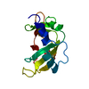
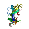
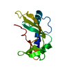
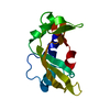
| Structure viewer | Molecule: MolmilJmol/JSmol |
|---|
- Downloads & links
-Download
| PDBx/mmCIF format | 1k5a.cif.gz | 37.1 KB | Display | PDBx/mmCIF format |
|---|---|---|---|---|
| PDB format | pdb1k5a.ent.gz | 24.6 KB | Display | PDB format |
| PDBx/mmJSON format | 1k5a.json.gz | Tree view | PDBx/mmJSON format | |
| Others |  Other downloads Other downloads |
-Validation report
| Arichive directory | https://data.pdbj.org/pub/pdb/validation_reports/k5/1k5aftp://data.pdbj.org/pub/pdb/validation_reports/k5/1k5a | HTTPS FTP |
|---|
-Related structure data
| Related structure data | 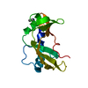 1k58C 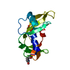 1k59C 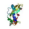 1k5bC 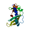 2angS S: Starting model for refinement C: citing same article ( |
|---|---|
| Similar structure data |


-Links
 PDBj
PDBj


- Assembly
| Deposited unit | 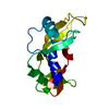
| ||||||||
|---|---|---|---|---|---|---|---|---|---|
| 1 |
| ||||||||
| Unit cell |
| ||||||||
| Components on special symmetry positions |
|
-Components
| #1: Protein | Mass: 14050.859 Da / Num. of mol.: 1 / Mutation: I119A,F120A Source method: isolated from a genetically manipulated source Source: (gene. exp.) Homo sapiens (human) / Production host:  References: UniProt: P03950, Hydrolases; Acting on ester bonds; Endoribonucleases producing 3'-phosphomonoesters |
|---|---|
| #2: Water | ChemComp-HOH /  Mass: 18.015 Da / Num. of mol.: 41 / Source method: isolated from a natural source / Formula: H2O Mass: 18.015 Da / Num. of mol.: 41 / Source method: isolated from a natural source / Formula: H2O |
| Has protein modification | Y |
-Experimental details
-Experiment
| Experiment | Method: X-RAY DIFFRACTION / Number of used crystals: 1 |
|---|
- Sample preparation
| Crystal | Density Matthews: 2.17 Å3/Da / Density % sol: 43.34 % | ||||||||||||||||||||||||||||||
|---|---|---|---|---|---|---|---|---|---|---|---|---|---|---|---|---|---|---|---|---|---|---|---|---|---|---|---|---|---|---|---|
| Crystal grow | Temperature: 289 K / Method: vapor diffusion, hanging drop / pH: 5.6 Details: ammonium actate, sodium citrate, PEG 4000, pH 5.6, VAPOR DIFFUSION, HANGING DROP, temperature 289K | ||||||||||||||||||||||||||||||
| Crystal grow | *PLUS Temperature: 16 ℃ / Method: vapor diffusion | ||||||||||||||||||||||||||||||
| Components of the solutions | *PLUS
|
-Data collection
| Diffraction | Mean temperature: 293 K |
|---|---|
| Diffraction source | Source: ROTATING ANODE / Type: SIEMENS / Wavelength: 1.5418 Å |
| Detector | Type: XENTRONICS / Detector: AREA DETECTOR / Date: Dec 20, 1996 |
| Radiation | Monochromator: GRAPHITE / Protocol: SINGLE WAVELENGTH / Monochromatic (M) / Laue (L): M / Scattering type: x-ray |
| Radiation wavelength | Wavelength: 1.5418 Å / Relative weight: 1 |
| Reflection | Resolution: 2.3→35 Å / Num. all: 5235 / Num. obs: 5235 / % possible obs: 88 % / Observed criterion σ(F): -3 / Observed criterion σ(I): -3 / Redundancy: 4.6 % / Biso Wilson estimate: 34.9 Å2 / Rsym value: 0.087 / Net I/σ(I): 15.5 |
| Reflection shell | Resolution: 2.3→2.4 Å / % possible all: 32 |
| Reflection | *PLUS Lowest resolution: 35 Å / % possible obs: 88 % / Num. measured all: 24206 / Rmerge(I) obs: 0.087 |
| Reflection shell | *PLUS % possible obs: 32 % / Mean I/σ(I) obs: 15.5 |
- Processing
| Software |
| ||||||||||||||||||||||||||||||||||||
|---|---|---|---|---|---|---|---|---|---|---|---|---|---|---|---|---|---|---|---|---|---|---|---|---|---|---|---|---|---|---|---|---|---|---|---|---|---|
| Refinement | Method to determine structure: MOLECULAR REPLACEMENT Starting model: PDN ENTRY 2ANG Resolution: 2.33→20 Å / Rfactor Rfree error: 0.017 / Data cutoff high absF: 10000000 / Data cutoff low absF: 0.001 / Isotropic thermal model: RESTRAINED / Cross valid method: THROUGHOUT / σ(F): 0 / Stereochemistry target values: Engh & Huber / Details: BULK SOLVENT MODEL USED
| ||||||||||||||||||||||||||||||||||||
| Displacement parameters | Biso mean: 32.1 Å2
| ||||||||||||||||||||||||||||||||||||
| Refine analyze |
| ||||||||||||||||||||||||||||||||||||
| Refinement step | Cycle: LAST / Resolution: 2.33→20 Å
| ||||||||||||||||||||||||||||||||||||
| Refine LS restraints |
| ||||||||||||||||||||||||||||||||||||
| LS refinement shell | Resolution: 2.33→2.48 Å / Rfactor Rfree error: 0.052 / Total num. of bins used: 6
| ||||||||||||||||||||||||||||||||||||
| Refinement | *PLUS Highest resolution: 2.3 Å / Lowest resolution: 20 Å / % reflection Rfree: 5 % | ||||||||||||||||||||||||||||||||||||
| Solvent computation | *PLUS | ||||||||||||||||||||||||||||||||||||
| Displacement parameters | *PLUS | ||||||||||||||||||||||||||||||||||||
| Refine LS restraints | *PLUS
| ||||||||||||||||||||||||||||||||||||
| LS refinement shell | *PLUS Rfactor obs: 0.294 |
