 ムービー
ムービー コントローラー


+ データを開く

- 基本情報
基本情報
| 登録情報 | データベース: PDB / ID: 1k4z | |||||||||
|---|---|---|---|---|---|---|---|---|---|---|
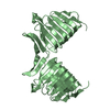
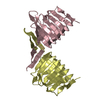
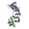
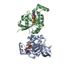
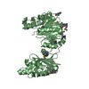
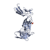
| タイトル | C-terminal Domain of Cyclase Associated Protein | |||||||||
 要素 要素 | Adenylyl Cyclase-Associated Protein | |||||||||
 キーワード キーワード | MEMBRANE PROTEIN / right-handed parallel beta-helix / intertwined dimer / actin-binding / New York SGX Research Center for Structural Genomics / NYSGXRC / Structural Genomics / PSI / Protein Structure Initiative | |||||||||
| 機能・相同性 |  機能・相同性情報 機能・相同性情報positive regulation of actin filament depolymerization / actin cortical patch / regulation of actin filament polymerization / actin filament severing / actin filament depolymerization / mating projection tip / positive regulation of Ras protein signal transduction / positive regulation of mitochondrial fission / adenylate cyclase binding / : ...positive regulation of actin filament depolymerization / actin cortical patch / regulation of actin filament polymerization / actin filament severing / actin filament depolymerization / mating projection tip / positive regulation of Ras protein signal transduction / positive regulation of mitochondrial fission / adenylate cyclase binding / : / Neutrophil degranulation / actin filament organization / actin binding / Ras protein signal transduction / identical protein binding / cytoplasm 類似検索 - 分子機能 | |||||||||
| 生物種 |  | |||||||||
| 手法 |  X線回折 / シンクロトロン / 多波長異常分散 / 解像度: 2.3 Å X線回折 / シンクロトロン / 多波長異常分散 / 解像度: 2.3 Å | |||||||||
 データ登録者 データ登録者 | Rozwarski, D.A. / Fedorov, A.A. / Dodatko, T. / Almo, S.C. / Burley, S.K. / New York SGX Research Center for Structural Genomics (NYSGXRC) | |||||||||
 引用 引用 | ジャーナル: Biochemistry / 年: 2004 タイトル: Crystal structure of the actin binding domain of the cyclase-associated protein 著者: Dodatko, T. / Fedorov, A.A. / Grynberg, M. / Patskovsky, Y. / Rozwarski, D.A. / Jaroszewski, L. / Aronoff-Spencer, E. / Kondraskina, E. / Irving, T. / Godzik, A. / Almo, S.C. #1: ジャーナル: J.Biol.Chem. / 年: 1996タイトル: Two Separate Functions Are Encoded by the Carboxyl-terminal Domains of the Yeast Cyclase-associated Protein and Its Mammalian Homologs. Dimerization and Actin Binding. 著者: Zelicof, A. / Protopopov, V. / David, D. / Lin, X.Y. / Lusgarten, V. / Gerst, J.E. | |||||||||
| 履歴 |
|
- 構造の表示
構造の表示
| 構造ビューア | 分子: MolmilJmol/JSmol |
|---|
- ダウンロードとリンク
-ダウンロード
| PDBx/mmCIF形式 | 1k4z.cif.gz | 77.3 KB | 表示 | PDBx/mmCIF形式 |
|---|---|---|---|---|
| PDB形式 | pdb1k4z.ent.gz | 57.2 KB | 表示 | PDB形式 |
| PDBx/mmJSON形式 | 1k4z.json.gz | ツリー表示 | PDBx/mmJSON形式 | |
| その他 |  その他のダウンロード その他のダウンロード |
-検証レポート
| 文書・要旨 | 1k4z_validation.pdf.gz | 370.1 KB | 表示 | wwPDB検証レポート |
|---|---|---|---|---|
| 文書・詳細版 | 1k4z_full_validation.pdf.gz | 372.1 KB | 表示 | |
| XML形式データ | 1k4z_validation.xml.gz | 7 KB | 表示 | |
| CIF形式データ | 1k4z_validation.cif.gz | 11.4 KB | 表示 | |
| アーカイブディレクトリ | https://data.pdbj.org/pub/pdb/validation_reports/k4/1k4zftp://data.pdbj.org/pub/pdb/validation_reports/k4/1k4z | HTTPS FTP |
-関連構造データ


-リンク
 PDBj
PDBj


- 集合体
集合体
| 登録構造単位 | 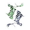
| ||||||||
|---|---|---|---|---|---|---|---|---|---|
| 1 | 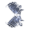
| ||||||||
| 2 | 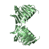
| ||||||||
| 単位格子 |
| ||||||||
| 詳細 | The biological assembly is an intertwined C-CAP dimer, which can be constructed using chain A and its symmetry partner generated by a crystallographic two-fold axis; or using chain B and its symmetry partner, generated by another crystallographic two-fold axis. |
-要素
| #1: タンパク質 | 分子量: 17540.641 Da / 分子数: 2 / 断片: C-TERMINAL DOMAIN / 由来タイプ: 組換発現 由来: (組換発現) 発現宿主:  #2: 水 | ChemComp-HOH / |  分子量: 18.015 Da / 分子数: 160 / 由来タイプ: 天然 / 式: H2O 分子量: 18.015 Da / 分子数: 160 / 由来タイプ: 天然 / 式: H2OHas protein modification | Y | |
|---|
-実験情報
-実験
| 実験 | 手法: X線回折 / 使用した結晶の数: 1 |
|---|
- 試料調製
試料調製
| 結晶 | マシュー密度: 2.827 Å3/Da / 溶媒含有率: 56.53 % | ||||||||||||||||||||||||||||||
|---|---|---|---|---|---|---|---|---|---|---|---|---|---|---|---|---|---|---|---|---|---|---|---|---|---|---|---|---|---|---|---|
| 結晶化 | 温度: 298 K / 手法: 蒸気拡散法, ハンギングドロップ法 / pH: 7.6 詳細: PEG 4000, lithium sulfate, HEPES, pH 7.6, VAPOR DIFFUSION, HANGING DROP, temperature 298.0K | ||||||||||||||||||||||||||||||
| 結晶化 | *PLUS 温度: 16 ℃ / 手法: 蒸気拡散法, シッティングドロップ法 / pH: 8.5 | ||||||||||||||||||||||||||||||
| 溶液の組成 | *PLUS
|
-データ収集
| 回折 | 平均測定温度: 103 K |
|---|---|
| 放射光源 | 由来: シンクロトロン / サイト: NSLS  / ビームライン: X9B / 波長: 1.04 Å / ビームライン: X9B / 波長: 1.04 Å |
| 検出器 | タイプ: MARRESEARCH / 検出器: IMAGE PLATE / 日付: 1998年8月30日 |
| 放射 | プロトコル: SINGLE WAVELENGTH / 単色(M)・ラウエ(L): M / 散乱光タイプ: x-ray |
| 放射波長 | 波長: 1.04 Å / 相対比: 1 |
| 反射 | 解像度: 2.3→10 Å / Num. all: 17249 / Num. obs: 17249 / % possible obs: 97.8 % / Observed criterion σ(F): 0 / Observed criterion σ(I): 0 / 冗長度: 6.1 % / Biso Wilson estimate: 37 Å2 / Rmerge(I) obs: 0.038 / Net I/σ(I): 49.7 |
| 反射 シェル | 解像度: 2.3→2.44 Å / 冗長度: 5 % / Rmerge(I) obs: 0.106 / Mean I/σ(I) obs: 16.4 / Num. unique all: 2732 / % possible all: 92.9 |
| 反射 | *PLUS 最高解像度: 2.3 Å / 最低解像度: 10 Å / Num. obs: 17479 / % possible obs: 97.3 % / Num. measured all: 105428 |
| 反射 シェル | *PLUS 最低解像度: 2.38 Å / % possible obs: 91.1 % / Num. unique obs: 1564 / Num. measured obs: 7805 / Rmerge(I) obs: 0.1 / Mean I/σ(I) obs: 18.1 |
- 解析
解析
| ソフトウェア |
| ||||||||||||||||||||||||||||||||||||
|---|---|---|---|---|---|---|---|---|---|---|---|---|---|---|---|---|---|---|---|---|---|---|---|---|---|---|---|---|---|---|---|---|---|---|---|---|---|
| 精密化 | 構造決定の手法: 多波長異常分散 / 解像度: 2.3→9.99 Å / Rfactor Rfree error: 0.006 / 交差検証法: THROUGHOUT / σ(F): 0 / 立体化学のターゲット値: Engh & Huber
| ||||||||||||||||||||||||||||||||||||
| 溶媒の処理 | 溶媒モデル: flat model / Bsol: 44.52 Å2 / ksol: 0.429148 e/Å3 | ||||||||||||||||||||||||||||||||||||
| 原子変位パラメータ | Biso mean: 32.3 Å2 | ||||||||||||||||||||||||||||||||||||
| Refine analyze |
| ||||||||||||||||||||||||||||||||||||
| 精密化ステップ | サイクル: LAST / 解像度: 2.3→9.99 Å
| ||||||||||||||||||||||||||||||||||||
| 拘束条件 |
| ||||||||||||||||||||||||||||||||||||
| Refine LS restraints NCS | NCS model details: restraints | ||||||||||||||||||||||||||||||||||||
| LS精密化 シェル | 解像度: 2.3→2.44 Å / Rfactor Rfree error: 0.018 / Total num. of bins used: 6
| ||||||||||||||||||||||||||||||||||||
| Xplor file |
| ||||||||||||||||||||||||||||||||||||
| 精密化 | *PLUS σ(F): 0 / % reflection Rfree: 10 % / Rfactor obs: 0.207 | ||||||||||||||||||||||||||||||||||||
| 溶媒の処理 | *PLUS | ||||||||||||||||||||||||||||||||||||
| 原子変位パラメータ | *PLUS Biso mean: 32.3 Å2 | ||||||||||||||||||||||||||||||||||||
| 拘束条件 | *PLUS
| ||||||||||||||||||||||||||||||||||||
| LS精密化 シェル | *PLUS % reflection Rfree: 9.3 % / Rfactor Rwork: 0.224 / Rfactor obs: 0.224 |
