 Movie
Movie Controller

[English] 日本語
 Yorodumi
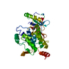
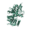
- PDB-1k3x: Crystal structure of a trapped reaction intermediate of the DNA r... -
+ Open data

- Basic information
| Entry | Database: PDB / ID: 1k3x | ||||||
|---|---|---|---|---|---|---|---|
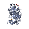
| Title | Crystal structure of a trapped reaction intermediate of the DNA repair enzyme Endonuclease VIII with Brominated-DNA | ||||||
 Components Components |
| ||||||
 Keywords Keywords | HYDROLASE/DNA / HYDROLASE-DNA complex | ||||||
| Function / homology |  Function and homology information Function and homology informationoxidized pyrimidine nucleobase lesion DNA N-glycosylase activity / Hydrolases; Glycosylases; Hydrolysing N-glycosyl compounds / DNA-(apurinic or apyrimidinic site) endonuclease activity / class I DNA-(apurinic or apyrimidinic site) endonuclease activity / DNA-(apurinic or apyrimidinic site) lyase / base-excision repair / damaged DNA binding / zinc ion binding Similarity search - Function | ||||||
| Biological species |  | ||||||
| Method |  X-RAY DIFFRACTION / SYNCHROTRON / AB INITIO PHASING / Resolution: 1.25 Å X-RAY DIFFRACTION / SYNCHROTRON / AB INITIO PHASING / Resolution: 1.25 Å | ||||||
 Authors Authors | Golan, G. / Zharkov, D.O. / Gilboa, R. / Fernandes, A.S. / Kycia, J.H. / Gerchman, S.E. / Rieger, R.A. / Grollman, A.P. / Shoham, G. | ||||||
 Citation Citation | Journal: EMBO J. / Year: 2002 Title: Structural analysis of an Escherichia coli endonuclease VIII covalent reaction intermediate. Authors: Zharkov, D.O. / Golan, G. / Gilboa, R. / Fernandes, A.S. / Gerchman, S.E. / Kycia, J.H. / Rieger, R.A. / Grollman, A.P. / Shoham, G. | ||||||
| History |
|
- Structure visualization
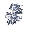
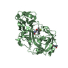
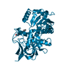
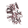
| Structure viewer | Molecule: MolmilJmol/JSmol |
|---|
- Downloads & links
-Download
| PDBx/mmCIF format | 1k3x.cif.gz | 168.4 KB | Display | PDBx/mmCIF format |
|---|---|---|---|---|
| PDB format | pdb1k3x.ent.gz | 127.2 KB | Display | PDB format |
| PDBx/mmJSON format | 1k3x.json.gz | Tree view | PDBx/mmJSON format | |
| Others |  Other downloads Other downloads |
-Validation report
| Arichive directory | https://data.pdbj.org/pub/pdb/validation_reports/k3/1k3xftp://data.pdbj.org/pub/pdb/validation_reports/k3/1k3x | HTTPS FTP |
|---|
-Related structure data
| Related structure data |  1k3wSC S: Starting model for refinement C: citing same article ( |
|---|---|
| Similar structure data |


-Links
 PDBj
PDBj


- Assembly
| Deposited unit | 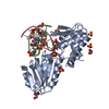
| ||||||||||||
|---|---|---|---|---|---|---|---|---|---|---|---|---|---|
| 1 |
| ||||||||||||
| Unit cell |
| ||||||||||||
| Components on special symmetry positions |
|
-Components
-DNA chain , 2 types, 2 molecules BC
| #1: DNA chain | Mass: 4218.051 Da / Num. of mol.: 1 / Source method: obtained synthetically |
|---|---|
| #2: DNA chain | Mass: 3863.531 Da / Num. of mol.: 1 / Source method: obtained synthetically |
-Protein , 1 types, 1 molecules A
| #3: Protein | Mass: 29814.994 Da / Num. of mol.: 1 Source method: isolated from a genetically manipulated source Source: (gene. exp.) References: UniProt: P50465, Hydrolases; Glycosylases; Hydrolysing N-glycosyl compounds |
|---|
-Non-polymers , 4 types, 498 molecules 


| #4: Chemical | ChemComp-ZN /  Mass: 65.409 Da / Num. of mol.: 1 / Source method: obtained synthetically / Formula: Zn Mass: 65.409 Da / Num. of mol.: 1 / Source method: obtained synthetically / Formula: Zn | ||||
|---|---|---|---|---|---|
| #5: Chemical | ChemComp-SO4 /  Mass: 96.063 Da / Num. of mol.: 5 / Source method: obtained synthetically / Formula: SO4 Mass: 96.063 Da / Num. of mol.: 5 / Source method: obtained synthetically / Formula: SO4#6: Chemical | ChemComp-GOL /  Mass: 92.094 Da / Num. of mol.: 4 / Source method: obtained synthetically / Formula: C3H8O3 Mass: 92.094 Da / Num. of mol.: 4 / Source method: obtained synthetically / Formula: C3H8O3#7: Water | ChemComp-HOH / | Mass: 18.015 Da / Num. of mol.: 488 / Source method: isolated from a natural source / Formula: H2O |
-Details
| Has protein modification | Y |
|---|
-Experimental details
-Experiment
| Experiment | Method: X-RAY DIFFRACTION / Number of used crystals: 1 |
|---|
- Sample preparation
| Crystal | Density Matthews: 2.99 Å3/Da / Density % sol: 61.11 % | |||||||||||||||||||||||||||||||||||||||||||||||||
|---|---|---|---|---|---|---|---|---|---|---|---|---|---|---|---|---|---|---|---|---|---|---|---|---|---|---|---|---|---|---|---|---|---|---|---|---|---|---|---|---|---|---|---|---|---|---|---|---|---|---|
| Crystal grow | Temperature: 288 K / Method: vapor diffusion, hanging drop / pH: 6.5 Details: 1.8M Ammonium-Sulfate, 0.1M Sodium-citrate pH 6.5, VAPOR DIFFUSION, HANGING DROP, temperature 288K | |||||||||||||||||||||||||||||||||||||||||||||||||
| Components of the solutions |
| |||||||||||||||||||||||||||||||||||||||||||||||||
| Crystal grow | *PLUS Temperature: 15 ℃ / Method: vapor diffusion / PH range low: 5 / PH range high: 4.6 | |||||||||||||||||||||||||||||||||||||||||||||||||
| Components of the solutions | *PLUS
|
-Data collection
| Diffraction | Mean temperature: 100 K |
|---|---|
| Diffraction source | Source: SYNCHROTRON / Site: NSLS  / Beamline: X25 / Wavelength: 0.92 / Beamline: X25 / Wavelength: 0.92 |
| Detector | Type: BRANDEIS - B4 / Detector: CCD / Date: May 29, 2001 |
| Radiation | Protocol: SINGLE WAVELENGTH / Monochromatic (M) / Laue (L): M / Scattering type: x-ray |
| Radiation wavelength | Wavelength: 0.92 Å / Relative weight: 1 |
| Reflection | Resolution: 1.25→40 Å / Num. obs: 131559 / % possible obs: 97.4 % / Redundancy: 7.5 % / Rsym value: 0.06 / Net I/σ(I): 9.7 |
| Reflection shell | Resolution: 1.25→1.27 Å / Redundancy: 7.5 % / Rsym value: 0.34 / % possible all: 91 |
| Reflection | *PLUS Num. measured all: 920114 / Rmerge(I) obs: 0.06 |
| Reflection shell | *PLUS % possible obs: 91 % / Rmerge(I) obs: 0.34 |
- Processing
| Software |
| |||||||||||||||||||||||||||||||||
|---|---|---|---|---|---|---|---|---|---|---|---|---|---|---|---|---|---|---|---|---|---|---|---|---|---|---|---|---|---|---|---|---|---|---|
| Refinement | Method to determine structure: AB INITIO PHASING Starting model: pdb entry 1k3w Resolution: 1.25→10 Å / Num. parameters: 26922 / Num. restraintsaints: 32739 / Cross valid method: FREE R / σ(F): 0 / Stereochemistry target values: ENGH & HUBER
| |||||||||||||||||||||||||||||||||
| Refine analyze | Luzzati coordinate error obs: 0.06 Å / Num. disordered residues: 8 / Occupancy sum hydrogen: 0 / Occupancy sum non hydrogen: 2941.52 | |||||||||||||||||||||||||||||||||
| Refinement step | Cycle: LAST / Resolution: 1.25→10 Å
| |||||||||||||||||||||||||||||||||
| Refine LS restraints |
| |||||||||||||||||||||||||||||||||
| LS refinement shell | Resolution: 1.25→1.27 Å
| |||||||||||||||||||||||||||||||||
| Software | *PLUS Name: SHELXL / Version: 97 / Classification: refinement | |||||||||||||||||||||||||||||||||
| Refinement | *PLUS Lowest resolution: 40 Å / % reflection Rfree: 10 % / Rfactor all: 0.149 / Rfactor Rfree: 0.1821 | |||||||||||||||||||||||||||||||||
| Solvent computation | *PLUS | |||||||||||||||||||||||||||||||||
| Displacement parameters | *PLUS | |||||||||||||||||||||||||||||||||
| Refine LS restraints | *PLUS
|
