 Movie
Movie Controller

[English] 日本語
 Yorodumi
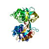
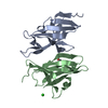
- PDB-1k3w: Crystal structure of a trapped reaction intermediate of the DNA R... -
+ Open data

- Basic information
| Entry | Database: PDB / ID: 1k3w | ||||||
|---|---|---|---|---|---|---|---|
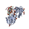
| Title | Crystal structure of a trapped reaction intermediate of the DNA Repair Enzyme Endonuclease VIII with DNA | ||||||
 Components Components |
| ||||||
 Keywords Keywords | HYDROLASE/DNA / HYDROLASE-DNA complex | ||||||
| Function / homology |  Function and homology information Function and homology informationoxidized pyrimidine nucleobase lesion DNA N-glycosylase activity / Hydrolases; Glycosylases; Hydrolysing N-glycosyl compounds / DNA-(apurinic or apyrimidinic site) endonuclease activity / class I DNA-(apurinic or apyrimidinic site) endonuclease activity / DNA-(apurinic or apyrimidinic site) lyase / base-excision repair / damaged DNA binding / zinc ion binding Similarity search - Function | ||||||
| Biological species |  | ||||||
| Method |  X-RAY DIFFRACTION / SYNCHROTRON / Resolution: 1.42 Å X-RAY DIFFRACTION / SYNCHROTRON / Resolution: 1.42 Å | ||||||
 Authors Authors | Golan, G. / Zharkov, D.O. / Gilboa, R. / Fernandes, A.S. / Kycia, J.H. / Gerchman, S.E. / Rieger, R.A. / Grollman, A.P. / Shoham, G. | ||||||
 Citation Citation | Journal: EMBO J. / Year: 2002 Title: Structural analysis of an Escherichia coli endonuclease VIII covalent reaction intermediate. Authors: Zharkov, D.O. / Golan, G. / Gilboa, R. / Fernandes, A.S. / Gerchman, S.E. / Kycia, J.H. / Rieger, R.A. / Grollman, A.P. / Shoham, G. | ||||||
| History |
|
- Structure visualization
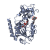
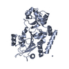
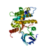
| Structure viewer | Molecule: MolmilJmol/JSmol |
|---|
- Downloads & links
-Download
| PDBx/mmCIF format | 1k3w.cif.gz | 159.5 KB | Display | PDBx/mmCIF format |
|---|---|---|---|---|
| PDB format | pdb1k3w.ent.gz | 119.7 KB | Display | PDB format |
| PDBx/mmJSON format | 1k3w.json.gz | Tree view | PDBx/mmJSON format | |
| Others |  Other downloads Other downloads |
-Validation report
| Arichive directory | https://data.pdbj.org/pub/pdb/validation_reports/k3/1k3wftp://data.pdbj.org/pub/pdb/validation_reports/k3/1k3w | HTTPS FTP |
|---|
-Related structure data


-Links
 PDBj
PDBj


- Assembly
| Deposited unit | 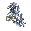
| ||||||||||||
|---|---|---|---|---|---|---|---|---|---|---|---|---|---|
| 1 |
| ||||||||||||
| Unit cell |
| ||||||||||||
| Components on special symmetry positions |
|
-Components
-DNA chain , 2 types, 2 molecules BC
| #1: DNA chain | Mass: 3958.571 Da / Num. of mol.: 1 / Source method: obtained synthetically |
|---|---|
| #2: DNA chain | Mass: 3863.531 Da / Num. of mol.: 1 / Source method: obtained synthetically |
-Protein , 1 types, 1 molecules A
| #3: Protein | Mass: 29814.994 Da / Num. of mol.: 1 Source method: isolated from a genetically manipulated source Source: (gene. exp.) References: UniProt: P50465, Hydrolases; Glycosylases; Hydrolysing N-glycosyl compounds |
|---|
-Non-polymers , 3 types, 401 molecules 


| #4: Chemical | ChemComp-ZN /  Mass: 65.409 Da / Num. of mol.: 1 / Source method: obtained synthetically / Formula: Zn Mass: 65.409 Da / Num. of mol.: 1 / Source method: obtained synthetically / Formula: Zn | ||
|---|---|---|---|
| #5: Chemical |  Mass: 96.063 Da / Num. of mol.: 3 / Source method: obtained synthetically / Formula: SO4 Mass: 96.063 Da / Num. of mol.: 3 / Source method: obtained synthetically / Formula: SO4#6: Water | ChemComp-HOH / | Mass: 18.015 Da / Num. of mol.: 397 / Source method: isolated from a natural source / Formula: H2O |
-Details
| Has protein modification | Y |
|---|
-Experimental details
-Experiment
| Experiment | Method: X-RAY DIFFRACTION / Number of used crystals: 1 |
|---|
- Sample preparation
| Crystal | Density Matthews: 3.03 Å3/Da / Density % sol: 60.79 % | |||||||||||||||||||||||||||||||||||||||||||||||||
|---|---|---|---|---|---|---|---|---|---|---|---|---|---|---|---|---|---|---|---|---|---|---|---|---|---|---|---|---|---|---|---|---|---|---|---|---|---|---|---|---|---|---|---|---|---|---|---|---|---|---|
| Crystal grow | Temperature: 288 K / Method: vapor diffusion, hanging drop / pH: 6.5 Details: 1.8M Ammonium-sulfate, 0.1M sodium-citrate pH 6.5, VAPOR DIFFUSION, HANGING DROP, temperature 288K | |||||||||||||||||||||||||||||||||||||||||||||||||
| Crystal grow | *PLUS Temperature: 15 ℃ / Method: vapor diffusion / PH range low: 5 / PH range high: 4.6 | |||||||||||||||||||||||||||||||||||||||||||||||||
| Components of the solutions | *PLUS
|
-Data collection
| Diffraction | Mean temperature: 100 K |
|---|---|
| Diffraction source | Source: SYNCHROTRON / Site: NSLS  / Beamline: X26C / Wavelength: 1.1 / Beamline: X26C / Wavelength: 1.1 |
| Detector | Type: ADSC QUANTUM 4 / Detector: CCD / Date: Feb 26, 2001 |
| Radiation | Protocol: SINGLE WAVELENGTH / Monochromatic (M) / Laue (L): M / Scattering type: x-ray |
| Radiation wavelength | Wavelength: 1.1 Å / Relative weight: 1 |
| Reflection | Resolution: 1.42→40 Å / Num. obs: 86586 / % possible obs: 95.2 % / Redundancy: 5.5 % / Rsym value: 0.083 / Net I/σ(I): 8.9 |
| Reflection shell | Resolution: 1.42→1.44 Å / Redundancy: 2 % / Num. unique all: 3191 / Rsym value: 0.375 / % possible all: 71.4 |
| Reflection | *PLUS Num. measured all: 471081 / Rmerge(I) obs: 0.08 |
| Reflection shell | *PLUS % possible obs: 71.4 % / Rmerge(I) obs: 0.375 |
- Processing
| Software |
| |||||||||||||||||||||||||||||||||
|---|---|---|---|---|---|---|---|---|---|---|---|---|---|---|---|---|---|---|---|---|---|---|---|---|---|---|---|---|---|---|---|---|---|---|
| Refinement | Starting model: initial MODEL OF BR-URACIL-CONTAINING DNA COMPLEXED WITH ENDONUCLEASE VIII AT 2.4A RESOLUTION Resolution: 1.42→10 Å / Num. parameters: 25384 / Num. restraintsaints: 31207 / Cross valid method: FREE R / σ(F): 0 / Stereochemistry target values: ENGH & HUBER
| |||||||||||||||||||||||||||||||||
| Solvent computation | Solvent model: MOEWS & KRETSINGER, J.MOL.BIOL.91(1973)201-228 | |||||||||||||||||||||||||||||||||
| Refine analyze | Num. disordered residues: 338 / Occupancy sum hydrogen: 0 / Occupancy sum non hydrogen: 2830.41 | |||||||||||||||||||||||||||||||||
| Refinement step | Cycle: LAST / Resolution: 1.42→10 Å
| |||||||||||||||||||||||||||||||||
| Refine LS restraints |
| |||||||||||||||||||||||||||||||||
| LS refinement shell | Resolution: 1.42→1.44 Å
| |||||||||||||||||||||||||||||||||
| Software | *PLUS Name: SHELXL / Version: 97 / Classification: refinement | |||||||||||||||||||||||||||||||||
| Refinement | *PLUS Lowest resolution: 40 Å / % reflection Rfree: 10 % / Rfactor all: 0.165 / Rfactor Rfree: 0.2026 / Rfactor Rwork: 0.1646 | |||||||||||||||||||||||||||||||||
| Solvent computation | *PLUS | |||||||||||||||||||||||||||||||||
| Displacement parameters | *PLUS | |||||||||||||||||||||||||||||||||
| Refine LS restraints | *PLUS
| |||||||||||||||||||||||||||||||||
| LS refinement shell | *PLUS Rfactor Rfree: 0.202 / Rfactor Rwork: 0.164 |
