 Movie
Movie Controller

[English] 日本語
 Yorodumi
- PDB-1k2b: Combining Mutations in HIV-1 Protease to Understand Mechanisms of... -
+ Open data

- Basic information
| Entry | Database: PDB / ID: 1k2b | ||||||
|---|---|---|---|---|---|---|---|
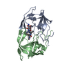
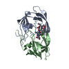
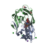
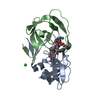
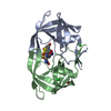
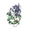
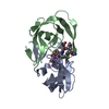
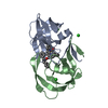
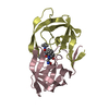
| Title | Combining Mutations in HIV-1 Protease to Understand Mechanisms of Resistance | ||||||
 Components Components | PROTEASE RETROPEPSIN | ||||||
 Keywords Keywords | HYDROLASE/HYDROLASE INHIBITOR / HIV-1 PROTEASE / HYDROLASE-HYDROLASE INHIBITOR COMPLEX | ||||||
| Function / homology |  Function and homology information Function and homology informationHIV-1 retropepsin / symbiont-mediated activation of host apoptosis / retroviral ribonuclease H / exoribonuclease H / exoribonuclease H activity / host multivesicular body / DNA integration / viral genome integration into host DNA / RNA-directed DNA polymerase / establishment of integrated proviral latency ...HIV-1 retropepsin / symbiont-mediated activation of host apoptosis / retroviral ribonuclease H / exoribonuclease H / exoribonuclease H activity / host multivesicular body / DNA integration / viral genome integration into host DNA / RNA-directed DNA polymerase / establishment of integrated proviral latency / viral penetration into host nucleus / RNA stem-loop binding / RNA-directed DNA polymerase activity / RNA-DNA hybrid ribonuclease activity / Transferases; Transferring phosphorus-containing groups; Nucleotidyltransferases / host cell / viral nucleocapsid / DNA recombination / DNA-directed DNA polymerase / aspartic-type endopeptidase activity / Hydrolases; Acting on ester bonds / DNA-directed DNA polymerase activity / symbiont-mediated suppression of host gene expression / viral translational frameshifting / lipid binding / symbiont entry into host cell / host cell nucleus / host cell plasma membrane / virion membrane / structural molecule activity / proteolysis / DNA binding / zinc ion binding / membrane Similarity search - Function | ||||||
| Biological species |   Human immunodeficiency virus 1 Human immunodeficiency virus 1 | ||||||
| Method |  X-RAY DIFFRACTION / SYNCHROTRON / MOLECULAR REPLACEMENT / Resolution: 1.7 Å X-RAY DIFFRACTION / SYNCHROTRON / MOLECULAR REPLACEMENT / Resolution: 1.7 Å | ||||||
 Authors Authors | Mahalingam, B. / Boross, P. / Wang, Y.-F. / Louis, J.M. / Fischer, C. / Tozser, J. / W Harrison, R. / Weber, I.T. | ||||||
 Citation Citation | Journal: Proteins / Year: 2002 Title: Combining mutations in HIV-1 protease to understand mechanisms of resistance. Authors: Mahalingam, B. / Boross, P. / Wang, Y.F. / Louis, J.M. / Fischer, C.C. / Tozser, J. / Harrison, R.W. / Weber, I.T. | ||||||
| History |
|
- Structure visualization
| Structure viewer | Molecule: MolmilJmol/JSmol |
|---|
- Downloads & links
-Download
| PDBx/mmCIF format | 1k2b.cif.gz | 56.6 KB | Display | PDBx/mmCIF format |
|---|---|---|---|---|
| PDB format | pdb1k2b.ent.gz | 40 KB | Display | PDB format |
| PDBx/mmJSON format | 1k2b.json.gz | Tree view | PDBx/mmJSON format | |
| Others |  Other downloads Other downloads |
-Validation report
| Summary document | 1k2b_validation.pdf.gz | 974.8 KB | Display | wwPDB validaton report |
|---|---|---|---|---|
| Full document | 1k2b_full_validation.pdf.gz | 980.6 KB | Display | |
| Data in XML | 1k2b_validation.xml.gz | 12.2 KB | Display | |
| Data in CIF | 1k2b_validation.cif.gz | 16.3 KB | Display | |
| Arichive directory | https://data.pdbj.org/pub/pdb/validation_reports/k2/1k2bftp://data.pdbj.org/pub/pdb/validation_reports/k2/1k2b | HTTPS FTP |
-Related structure data
| Related structure data |  1k1tC  1k1uC  1k2cC  1dazS S: Starting model for refinement C: citing same article ( |
|---|---|
| Similar structure data |

-Links
 PDBj
PDBj

- Assembly
| Deposited unit | 
| ||||||||
|---|---|---|---|---|---|---|---|---|---|
| 1 |
| ||||||||
| Unit cell |
|
-Components
| #1: Protein | Mass: 10759.699 Da / Num. of mol.: 2 / Mutation: Q7K, l33I, L63I, C67A, C95A, N88D, L90M Source method: isolated from a genetically manipulated source Source: (gene. exp.) Human immunodeficiency virus 1 / Genus: Lentivirus / Production host:  #2: Chemical | ChemComp-0Q4 / |   Type: peptide-like, Peptide-like / Class: Inhibitor / Mass: 833.053 Da / Num. of mol.: 1 / Source method: obtained synthetically / Formula: C40H70N11O8 Type: peptide-like, Peptide-like / Class: Inhibitor / Mass: 833.053 Da / Num. of mol.: 1 / Source method: obtained synthetically / Formula: C40H70N11O8References: N-[(2R)-2-({N~5~-[amino(iminio)methyl]-L-ornithyl-L-valyl}amino)-4-methylpentyl]-L-phenylalanyl-L-alpha-glutamyl- L-alanyl-L-norleucinamide #3: Water | ChemComp-HOH / |  Mass: 18.015 Da / Num. of mol.: 128 / Source method: isolated from a natural source / Formula: H2O Mass: 18.015 Da / Num. of mol.: 128 / Source method: isolated from a natural source / Formula: H2ONonpolymer details | THE INHIBITOR 0Q4 WAS CHEMICALLY SYNTHESIZED AND IS ANALOGOUS TO THE CA-P2 PROCESSING SITE ...THE INHIBITOR 0Q4 WAS CHEMICALLY | Sequence details | MUTATIONS Q7K, L33I, L63I, C67A, C95A, HAVE BEEN MADE TO STABILIZE THE PROTEASE FROM ...MUTATIONS Q7K, L33I, L63I, C67A, C95A, HAVE BEEN MADE TO STABILIZE THE PROTEASE FROM AUTOPROTEO | |
|---|
-Experimental details
-Experiment
| Experiment | Method: X-RAY DIFFRACTION / Number of used crystals: 1 |
|---|
- Sample preparation
| Crystal | Density Matthews: 1.99 Å3/Da / Density % sol: 38.08 % | ||||||||||||||||||||||||||||||||||||||||||
|---|---|---|---|---|---|---|---|---|---|---|---|---|---|---|---|---|---|---|---|---|---|---|---|---|---|---|---|---|---|---|---|---|---|---|---|---|---|---|---|---|---|---|---|
| Crystal grow | Temperature: 298 K / Method: vapor diffusion, hanging drop Details: 20-50% Saturated Ammonium Sulphate, 10% DMSO, 0.25M citrate/0.5M phosphate buffer, VAPOR DIFFUSION, HANGING DROP, temperature 298K | ||||||||||||||||||||||||||||||||||||||||||
| Crystal grow | *PLUS PH range low: 6.5 / PH range high: 5 | ||||||||||||||||||||||||||||||||||||||||||
| Components of the solutions | *PLUS
|
-Data collection
| Diffraction | Mean temperature: 90 K |
|---|---|
| Diffraction source | Source: SYNCHROTRON / Site: NSLS  / Beamline: X12B / Wavelength: 1.037 Å / Beamline: X12B / Wavelength: 1.037 Å |
| Detector | Type: ADSC QUANTUM 4 / Detector: CCD / Date: Oct 8, 1999 |
| Radiation | Monochromator: Double-crystal, fixed-exit Si-III / Protocol: SINGLE WAVELENGTH / Monochromatic (M) / Laue (L): M / Scattering type: x-ray |
| Radiation wavelength | Wavelength: 1.037 Å / Relative weight: 1 |
| Reflection | Resolution: 1.7→25 Å / Num. all: 20000 / Num. obs: 20000 / % possible obs: 99 % / Observed criterion σ(F): 2 / Observed criterion σ(I): 2 / Redundancy: 4.1 % / Rmerge(I) obs: 0.042 / Net I/σ(I): 10.8 |
| Reflection shell | Resolution: 1.7→1.76 Å / Rmerge(I) obs: 0.248 / Mean I/σ(I) obs: 5.1 / Num. unique all: 1958 / % possible all: 98.4 |
| Reflection | *PLUS Lowest resolution: 8 Å / % possible obs: 99 % / Rmerge(I) obs: 0.042 |
| Reflection shell | *PLUS Rmerge(I) obs: 0.248 |
- Processing
| Software |
| ||||||||||||||||||||||||
|---|---|---|---|---|---|---|---|---|---|---|---|---|---|---|---|---|---|---|---|---|---|---|---|---|---|
| Refinement | Method to determine structure: MOLECULAR REPLACEMENT Starting model: PDB ENTRY 1DAZ Resolution: 1.7→8 Å / Isotropic thermal model: Isotropic / Cross valid method: FREE R / σ(F): 2 / Stereochemistry target values: ENGH AND HUBER
| ||||||||||||||||||||||||
| Refinement step | Cycle: LAST / Resolution: 1.7→8 Å
| ||||||||||||||||||||||||
| Refine LS restraints |
| ||||||||||||||||||||||||
| LS refinement shell | Resolution: 1.7→1.76 Å
| ||||||||||||||||||||||||
| Refinement | *PLUS Rfactor Rfree: 0.275 / Rfactor Rwork: 0.216 | ||||||||||||||||||||||||
| Solvent computation | *PLUS | ||||||||||||||||||||||||
| Displacement parameters | *PLUS | ||||||||||||||||||||||||
| Refine LS restraints | *PLUS
| ||||||||||||||||||||||||
| LS refinement shell | *PLUS Highest resolution: 1.7 Å / Rfactor Rfree: 0.3784 / Rfactor Rwork: 0.3196 |
