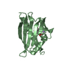
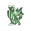
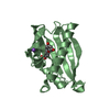
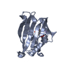
10 lowest energy structures consistent with experimental distance and dihedral angle restraints
Representative
Model #1
lowest energy
-
Components
#1: RNA chain
5'-R(*UP*AP*UP*AP*CP*UP*AP*AP*CP*AP*A)-3'
Mass: 3459.138 Da / Num. of mol.: 1 / Source method: obtained synthetically / Details: Yeast and mammalian consensus BPS sequence
#2: Protein
SF1-Boisoform / Splicing Factor 1
Mass: 14702.896 Da / Num. of mol.: 1 / Fragment: Residues 133-260, KH-QUA2 region Source method: isolated from a genetically manipulated source Source: (gene. exp.) Homo sapiens (human) / Plasmid: modified pET24d / Species (production host): Escherichia coli / Production host: Escherichia coli BL21(DE3) (bacteria) / Strain (production host): BL21 DE3 / References: UniProt: Q15637
-
Experimental details
-
Experiment
Experiment
Method: SOLUTION NMR
NMR experiment
Conditions-ID
Experiment-ID
Solution-ID
Type
1
1
2
3D 13C-separated NOESY
1
2
2
3D 13C/15N edited/filtered NOESY
1
3
3
3D 15N-separated NOESY
NMR details
Text: Backbone and side chain 1H, 15N and 13C resonances were assigned using standard triple resonance experiments. Distance restraints were derived from 13C- and 15N-edited 3D NOESY experiments. ...Text: Backbone and side chain 1H, 15N and 13C resonances were assigned using standard triple resonance experiments. Distance restraints were derived from 13C- and 15N-edited 3D NOESY experiments. Assignments for the RNA were obtained from 2D isotope-filtered experiments. Intermolecular distance restraints were measured in 3D 13C-edited/filtered experiments. Dihedral angle restraints for the backbone angle phi were derived from 3J(HN,Ha) coupling constants measured in an HNHA-J experiment, additional phi/psi restraints were derived from TALOS. Hydrogen bond restraints for secondary structure elements in the protein were defined from slowly exchanging amide protons, identified after exchange of the H2O buffer to D2O.
-
Sample preparation
Details
Solution-ID
Contents
Solvent system
1
1 mM U-15N,13C SF1 KH-QUA2/unlabeled BPS in 20 mM phosphate buffer NA pH 6.5, 50 mM NaCl, 2 mM DTT
90% H2O/10% D2O
2
1 mM U-15N,13C SF1 KH-QUA2/unlabeled BPS in 20 mM phosphate buffer NA pH 6.5, 50 mM NaCl, 2 mM DTT
100% D2O
3
1 mM U-15N SF1 KH-QUA2/unlabeled BPS in 20 mM phosphate buffer NA pH 6.5, 50 mM NaCl, 2 mM DTT
90% H2O/10% D2O
Sample conditions
Conditions-ID
Ionic strength
pH
Pressure (kPa)
Temperature (K)
1
50mMNaCl
6.5
ambient
295K
2
50mMNaCl
6.5
ambient
278K
Crystal grow
*PLUS
Method: other / Details: NMR
-
NMR measurement
Radiation
Protocol: SINGLE WAVELENGTH / Monochromatic (M) / Laue (L): M
Radiation wavelength
Relative weight: 1
NMR spectrometer
Type
Manufacturer
Model
Field strength (MHz)
Spectrometer-ID
Bruker DRX
Bruker
DRX
500
1
Bruker DRX
Bruker
DRX
600
2
Bruker DRX
Bruker
DRX
800
3
-
Processing
NMR software
Name
Version
Developer
Classification
XwinNMR
2.6
Bruker
collection
NMRPipe
1.8
F. Delaglio, S. Grzesiek, G. Vuister, G. Zhu, J. Pfeifer, & A. Bax
processing
XEASY
1.2
Ch. Bartels, T.-H. Xia, M. Billeter, P. Gntert and K. Wthrich
dataanalysis
ARIA/CNS
1
J. Linge, M. Nilges
structuresolution
ARIA/CNS
1
J. Linge, M. Nilges
refinement
Refinement
Method: restrained molecular dynamics, simulated annealing / Software ordinal: 1 Details: STRUCTURES WERE CALCULATED WITH A MIXED TORSION AND CARTESIAN ANGLE DYNAMICS PROTOCOL USING ARIA/CNS. A REDUCED RELAXATION MATRIX APPROACH WAS USED FOR THE NOE CALIBRATION. THE STRUCTURES ...Details: STRUCTURES WERE CALCULATED WITH A MIXED TORSION AND CARTESIAN ANGLE DYNAMICS PROTOCOL USING ARIA/CNS. A REDUCED RELAXATION MATRIX APPROACH WAS USED FOR THE NOE CALIBRATION. THE STRUCTURES ARE CURRENTLY BEING REFINED USING RESIDUAL DIPOLAR COUPLINGS AND WILL BE UPDATED IN THE FUTURE.
NMR representative
Selection criteria: lowest energy
NMR ensemble
Conformer selection criteria: 10 lowest energy structures consistent with experimental distance and dihedral angle restraints Conformers calculated total number: 100 / Conformers submitted total number: 10
+
About Yorodumi
-
News
-
Feb 9, 2022. New format data for meta-information of EMDB entries
New format data for meta-information of EMDB entries
Version 3 of the EMDB header file is now the official format.
The previous official version 1.9 will be removed from the archive.
In the structure databanks used in Yorodumi, some data are registered as the other names, "COVID-19 virus" and "2019-nCoV". Here are the details of the virus and the list of structure data.
Jan 31, 2019. EMDB accession codes are about to change! (news from PDBe EMDB page)
EMDB accession codes are about to change! (news from PDBe EMDB page)
The allocation of 4 digits for EMDB accession codes will soon come to an end. Whilst these codes will remain in use, new EMDB accession codes will include an additional digit and will expand incrementally as the available range of codes is exhausted. The current 4-digit format prefixed with “EMD-” (i.e. EMD-XXXX) will advance to a 5-digit format (i.e. EMD-XXXXX), and so on. It is currently estimated that the 4-digit codes will be depleted around Spring 2019, at which point the 5-digit format will come into force.
The EM Navigator/Yorodumi systems omit the EMD- prefix.
Related info.:Q: What is EMD? / ID/Accession-code notation in Yorodumi/EM Navigator
Yorodumi is a browser for structure data from EMDB, PDB, SASBDB, etc.
This page is also the successor to EM Navigator detail page, and also detail information page/front-end page for Omokage search.
The word "yorodu" (or yorozu) is an old Japanese word meaning "ten thousand". "mi" (miru) is to see.
Related info.:EMDB / PDB / SASBDB / Comparison of 3 databanks / Yorodumi Search / Aug 31, 2016. New EM Navigator & Yorodumi / Yorodumi Papers / Jmol/JSmol / Function and homology information / Changes in new EM Navigator and Yorodumi
 Movie
Movie Controller

 Yorodumi
 Open data
 Basic information
 Components
 Keywords
 Function and homology information
 Homo sapiens (human)
 Authors
 Citation
 Structure visualization
 Downloads & links
 Other downloads


 PDBj
PDBj


 Assembly

 Sample preparation
 Processing
 NMRPipe