 Movie
Movie Controller

[English] 日本語
 Yorodumi
- PDB-1jzb: Crystal Structure of Variant 2 Scorpion Toxin from Centruroides s... -
+ Open data

- Basic information
| Entry | Database: PDB / ID: 1jzb | ||||||
|---|---|---|---|---|---|---|---|
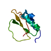
| Title | Crystal Structure of Variant 2 Scorpion Toxin from Centruroides sculpturatus Ewing | ||||||
 Components Components | NEUROTOXIN 2 | ||||||
 Keywords Keywords | TOXIN / scorpion toxin / noncrystallographic symmetry | ||||||
| Function / homology |  Function and homology information Function and homology informationsodium channel inhibitor activity / defense response / toxin activity / extracellular region Similarity search - Function | ||||||
| Biological species |  | ||||||
| Method |  X-RAY DIFFRACTION / MOLECULAR REPLACEMENT / Resolution: 2.81 Å X-RAY DIFFRACTION / MOLECULAR REPLACEMENT / Resolution: 2.81 Å | ||||||
 Authors Authors | Cook, W.J. / Zell, A. / Watt, D.D. / Ealick, S.E. | ||||||
 Citation Citation | Journal: Protein Sci. / Year: 2002 Title: Structure of variant 2 scorpion toxin from Centruroides sculpturatus Ewing. Authors: Cook, W.J. / Zell, A. / Watt, D.D. / Ealick, S.E. | ||||||
| History |
| ||||||
| Remark 999 | SEQUENCE THE AUTHORS MAINTAIN THAT THEIR SEQUENCE IS CORRECT. |
- Structure visualization
| Structure viewer | Molecule: MolmilJmol/JSmol |
|---|
- Downloads & links
-Download
| PDBx/mmCIF format | 1jzb.cif.gz | 19.1 KB | Display | PDBx/mmCIF format |
|---|---|---|---|---|
| PDB format | pdb1jzb.ent.gz | 14.5 KB | Display | PDB format |
| PDBx/mmJSON format | 1jzb.json.gz | Tree view | PDBx/mmJSON format | |
| Others |  Other downloads Other downloads |
-Validation report
| Summary document | 1jzb_validation.pdf.gz | 420.8 KB | Display | wwPDB validaton report |
|---|---|---|---|---|
| Full document | 1jzb_full_validation.pdf.gz | 423 KB | Display | |
| Data in XML | 1jzb_validation.xml.gz | 4.8 KB | Display | |
| Data in CIF | 1jzb_validation.cif.gz | 5.6 KB | Display | |
| Arichive directory | https://data.pdbj.org/pub/pdb/validation_reports/jz/1jzbftp://data.pdbj.org/pub/pdb/validation_reports/jz/1jzb | HTTPS FTP |
-Related structure data


-Links
 PDBj
PDBj

- Assembly
| Deposited unit | 
| ||||||||
|---|---|---|---|---|---|---|---|---|---|
| 1 |
| ||||||||
| Unit cell |
|
-Components
| #1: Protein | Mass: 7182.110 Da / Num. of mol.: 1 / Source method: isolated from a natural source / Source: (natural) |
|---|
-Experimental details
-Experiment
| Experiment | Method: X-RAY DIFFRACTION / Number of used crystals: 1 |
|---|
- Sample preparation
| Crystal | Density Matthews: 2.09 Å3/Da / Density % sol: 41.16 % | ||||||||||||||||||||||||
|---|---|---|---|---|---|---|---|---|---|---|---|---|---|---|---|---|---|---|---|---|---|---|---|---|---|
| Crystal grow | Temperature: 277 K / Method: vapor diffusion, hanging drop / pH: 9.2 Details: 2-methyl-2,4-pentanediol, CHES buffer, pH 9.2, VAPOR DIFFUSION, HANGING DROP, temperature 277K | ||||||||||||||||||||||||
| Crystal grow | *PLUS Temperature: 4 ℃ / Details: Ealick, S.E., (1984) J. Biol. Chem., 259, 12081. / PH range low: 9.8 / PH range high: 8.6 | ||||||||||||||||||||||||
| Components of the solutions | *PLUS
|
-Data collection
| Diffraction | Mean temperature: 296 K |
|---|---|
| Diffraction source | Source: SEALED TUBE / Type: OTHER / Wavelength: 1.5418 Å |
| Detector | Type: PICKER / Detector: DIFFRACTOMETER / Date: Jan 1, 1985 |
| Radiation | Monochromator: Ni FILTER / Protocol: SINGLE WAVELENGTH / Monochromatic (M) / Laue (L): M / Scattering type: x-ray |
| Radiation wavelength | Wavelength: 1.5418 Å / Relative weight: 1 |
| Reflection | Resolution: 2.8→100 Å / Num. all: 1609 / Num. obs: 1609 / % possible obs: 99.2 % / Observed criterion σ(F): 0 / Observed criterion σ(I): 0 |
| Reflection shell | Resolution: 2.8→2.98 Å / % possible all: 96.7 |
| Reflection | *PLUS Lowest resolution: 100 Å |
- Processing
| Software |
| ||||||||||||||||||||||||||||||||||||
|---|---|---|---|---|---|---|---|---|---|---|---|---|---|---|---|---|---|---|---|---|---|---|---|---|---|---|---|---|---|---|---|---|---|---|---|---|---|
| Refinement | Method to determine structure: MOLECULAR REPLACEMENT / Resolution: 2.81→42.26 Å / Rfactor Rfree error: 0.031 / Data cutoff high absF: 125210.84 / Data cutoff low absF: 0 / Isotropic thermal model: RESTRAINED / Cross valid method: THROUGHOUT / σ(F): 0 / Stereochemistry target values: Engh & Huber
| ||||||||||||||||||||||||||||||||||||
| Solvent computation | Solvent model: FLAT MODEL / Bsol: 122.39 Å2 / ksol: 0.582076 e/Å3 | ||||||||||||||||||||||||||||||||||||
| Displacement parameters | Biso mean: 17.8 Å2
| ||||||||||||||||||||||||||||||||||||
| Refine analyze |
| ||||||||||||||||||||||||||||||||||||
| Refinement step | Cycle: LAST / Resolution: 2.81→42.26 Å
| ||||||||||||||||||||||||||||||||||||
| Refine LS restraints |
| ||||||||||||||||||||||||||||||||||||
| LS refinement shell | Resolution: 2.8→2.98 Å / Rfactor Rfree error: 0.121 / Total num. of bins used: 6
| ||||||||||||||||||||||||||||||||||||
| Xplor file | Serial no: 1 / Param file: PROTEIN_REP.PARAM / Topol file: PROTEIN.TOP | ||||||||||||||||||||||||||||||||||||
| Software | *PLUS Name: CNS / Version: 1 / Classification: refinement | ||||||||||||||||||||||||||||||||||||
| Refinement | *PLUS Highest resolution: 2.8 Å / Lowest resolution: 50 Å / σ(F): 0 / % reflection Rfree: 5 % / Rfactor obs: 0.229 / Rfactor Rfree: 0.281 | ||||||||||||||||||||||||||||||||||||
| Solvent computation | *PLUS | ||||||||||||||||||||||||||||||||||||
| Displacement parameters | *PLUS Biso mean: 17.8 Å2 | ||||||||||||||||||||||||||||||||||||
| Refine LS restraints | *PLUS
| ||||||||||||||||||||||||||||||||||||
| LS refinement shell | *PLUS Rfactor Rfree: 0.283 / % reflection Rfree: 3.1 % / Rfactor Rwork: 0.283 / Num. reflection obs: 260 / Rfactor obs: 0.286 |
