 Movie
Movie Controller


+ Open data

- Basic information
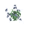
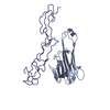
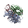
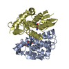
| Entry | Database: PDB / ID: 1jtz | ||||||
|---|---|---|---|---|---|---|---|
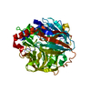
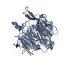
| Title | CRYSTAL STRUCTURE OF TRANCE/RANKL CYTOKINE. | ||||||
 Components Components | TUMOR NECROSIS FACTOR LIGAND SUPERFAMILY MEMBER 11 | ||||||
 Keywords Keywords | CYTOKINE / TUMOR NECROSIS FACTOR SUPERFAMILY MEMBER / JELLYROLL / BETA-SANDWICH | ||||||
| Function / homology |  Function and homology information Function and homology informationtooth eruption / positive regulation of fever generation by positive regulation of prostaglandin secretion / positive regulation of corticotropin-releasing hormone secretion / TNF receptor superfamily (TNFSF) members mediating non-canonical NF-kB pathway / osteoclast proliferation / TNFs bind their physiological receptors / positive regulation of osteoclast development / tumor necrosis factor receptor superfamily binding / Transcriptional and post-translational regulation of MITF-M expression and activity / TNFR2 non-canonical NF-kB pathway ...tooth eruption / positive regulation of fever generation by positive regulation of prostaglandin secretion / positive regulation of corticotropin-releasing hormone secretion / TNF receptor superfamily (TNFSF) members mediating non-canonical NF-kB pathway / osteoclast proliferation / TNFs bind their physiological receptors / positive regulation of osteoclast development / tumor necrosis factor receptor superfamily binding / Transcriptional and post-translational regulation of MITF-M expression and activity / TNFR2 non-canonical NF-kB pathway / osteoclast development / positive regulation of homotypic cell-cell adhesion / regulation of osteoclast differentiation / paracrine signaling / positive regulation of osteoclast differentiation / positive regulation of bone resorption / tumor necrosis factor receptor binding / mammary gland epithelial cell proliferation / positive regulation of extrinsic apoptotic signaling pathway / mammary gland alveolus development / positive regulation of intracellular signal transduction / monocyte chemotaxis / bone resorption / lymph node development / calcium ion homeostasis / cellular response to leukemia inhibitory factor / osteoclast differentiation / ossification / cytokine activity / animal organ morphogenesis / tumor necrosis factor-mediated signaling pathway / calcium-mediated signaling / positive regulation of non-canonical NF-kappaB signal transduction / bone development / positive regulation of JNK cascade / positive regulation of T cell activation / cytokine-mediated signaling pathway / positive regulation of MAPK cascade / positive regulation of ERK1 and ERK2 cascade / positive regulation of canonical NF-kappaB signal transduction / positive regulation of phosphatidylinositol 3-kinase/protein kinase B signal transduction / cell surface receptor signaling pathway / immune response / receptor ligand activity / positive regulation of gene expression / negative regulation of transcription by RNA polymerase II / positive regulation of transcription by RNA polymerase II / : / identical protein binding / plasma membrane / cytoplasm Similarity search - Function | ||||||
| Biological species |  | ||||||
| Method |  X-RAY DIFFRACTION / MOLECULAR REPLACEMENT / Resolution: 2.6 Å X-RAY DIFFRACTION / MOLECULAR REPLACEMENT / Resolution: 2.6 Å | ||||||
 Authors Authors | Nelson, C.A. / Fremont, D.H. | ||||||
 Citation Citation | Journal: J.Clin.Invest. / Year: 2001 Title: Crystal structure of the TRANCE/RANKL cytokine reveals determinants of receptor-ligand specificity Authors: Lam, J. / Nelson, C.A. / Ross, F.P. / Teitelbaum, S.L. / Fremont, D.H. | ||||||
| History |
|
- Structure visualization
| Structure viewer | Molecule: MolmilJmol/JSmol |
|---|
- Downloads & links
-Download
| PDBx/mmCIF format | 1jtz.cif.gz | 107 KB | Display | PDBx/mmCIF format |
|---|---|---|---|---|
| PDB format | pdb1jtz.ent.gz | 82.5 KB | Display | PDB format |
| PDBx/mmJSON format | 1jtz.json.gz | Tree view | PDBx/mmJSON format | |
| Others |  Other downloads Other downloads |
-Validation report
| Arichive directory | https://data.pdbj.org/pub/pdb/validation_reports/jt/1jtzftp://data.pdbj.org/pub/pdb/validation_reports/jt/1jtz | HTTPS FTP |
|---|
-Related structure data


-Links
 PDBj
PDBj
- Assembly
| Deposited unit | 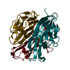
| |||||||||||||||||||||||||||||||||
|---|---|---|---|---|---|---|---|---|---|---|---|---|---|---|---|---|---|---|---|---|---|---|---|---|---|---|---|---|---|---|---|---|---|---|
| 1 |
| |||||||||||||||||||||||||||||||||
| Unit cell |
| |||||||||||||||||||||||||||||||||
| Noncrystallographic symmetry (NCS) | NCS domain:
| |||||||||||||||||||||||||||||||||
| Details | THE BIOLOGICAL ASSEMBLY IS A TRIMER WHICH COMPRISES THE ASYMETRIC UNIT. |
-Components
| #1: Protein | Mass: 19056.359 Da / Num. of mol.: 3 Fragment: C-TERMINAL RECEPTOR-BINDING ECTODOMAIN, Residues 156-316 Source method: isolated from a genetically manipulated source Source: (gene. exp.)  #2: Water | ChemComp-HOH / |  Mass: 18.015 Da / Num. of mol.: 147 / Source method: isolated from a natural source / Formula: H2O Mass: 18.015 Da / Num. of mol.: 147 / Source method: isolated from a natural source / Formula: H2O |
|---|
-Experimental details
-Experiment
| Experiment | Method: X-RAY DIFFRACTION / Number of used crystals: 1 |
|---|
- Sample preparation
| Crystal | Density Matthews: 2.33 Å3/Da / Density % sol: 46 % | ||||||||||||||||||||||||||||||||||||||||||
|---|---|---|---|---|---|---|---|---|---|---|---|---|---|---|---|---|---|---|---|---|---|---|---|---|---|---|---|---|---|---|---|---|---|---|---|---|---|---|---|---|---|---|---|
| Crystal grow | Temperature: 293 K / Method: vapor diffusion, hanging drop / pH: 7.5 Details: 11-14% PEG 4000, 16 mM calcium chloride, 80 mM Hepes, pH 7.5, VAPOR DIFFUSION, HANGING DROP, temperature 293K | ||||||||||||||||||||||||||||||||||||||||||
| Crystal grow | *PLUS Temperature: 20 ℃ / pH: 8 | ||||||||||||||||||||||||||||||||||||||||||
| Components of the solutions | *PLUS
|
-Data collection
| Diffraction | Mean temperature: 110 K |
|---|---|
| Diffraction source | Source: ROTATING ANODE / Type: RIGAKU RU200 / Wavelength: 1.5418 Å |
| Detector | Type: RIGAKU RAXIS IV / Detector: IMAGE PLATE / Date: Dec 4, 2000 / Details: YALE MIRRORS |
| Radiation | Protocol: SINGLE WAVELENGTH / Monochromatic (M) / Laue (L): M / Scattering type: x-ray |
| Radiation wavelength | Wavelength: 1.5418 Å / Relative weight: 1 |
| Reflection | Resolution: 2.6→20 Å / Num. all: 191355 / Num. obs: 16705 / % possible obs: 98.2 % / Observed criterion σ(F): 0 / Observed criterion σ(I): -3 / Redundancy: 11.5 % / Biso Wilson estimate: 70.2 Å2 / Rsym value: 0.059 / Net I/σ(I): 32.3 |
| Reflection shell | Resolution: 2.6→2.72 Å / Redundancy: 11.5 % / Mean I/σ(I) obs: 4.7 / Rsym value: 0.449 / % possible all: 100 |
| Reflection | *PLUS Num. measured all: 191355 / Rmerge F obs: 0.059 |
| Reflection shell | *PLUS % possible obs: 100 % / Rmerge(I) obs: 0.449 |
- Processing
| Software |
| ||||||||||||||||||||||||||||||||||||||||||||||||||||||||||||||||||
|---|---|---|---|---|---|---|---|---|---|---|---|---|---|---|---|---|---|---|---|---|---|---|---|---|---|---|---|---|---|---|---|---|---|---|---|---|---|---|---|---|---|---|---|---|---|---|---|---|---|---|---|---|---|---|---|---|---|---|---|---|---|---|---|---|---|---|---|
| Refinement | Method to determine structure: MOLECULAR REPLACEMENT Starting model: CHIMERA OF PDB ENTRIES 1TNR (CHAIN A) AND 1D4V (CHAIN B) Resolution: 2.6→20 Å / Rfactor Rfree error: 0.01 / Data cutoff high absF: 53633.89 / Data cutoff low absF: 0 / Isotropic thermal model: RESTRAINED / Cross valid method: THROUGHOUT / σ(F): 0 / Stereochemistry target values: MLF Details: The following residues were included in 5 groups NCS GROUP 1:162-171,180-223,235-244,252-263,269-316 NCS GROUP 2:172-179 NCS GROUP 3:224-233 NCS GROUP 4:245-251 NCS GROUP 5:264-268 NOTES: ...Details: The following residues were included in 5 groups NCS GROUP 1:162-171,180-223,235-244,252-263,269-316 NCS GROUP 2:172-179 NCS GROUP 3:224-233 NCS GROUP 4:245-251 NCS GROUP 5:264-268 NOTES: Residues included in groups 1-5 (RMS for monomer X versus monomer Y) is identical to groups 6-10 (RMS for monomer X versus monomer Z)
| ||||||||||||||||||||||||||||||||||||||||||||||||||||||||||||||||||
| Solvent computation | Solvent model: FLAT MODEL / Bsol: 36.7347 Å2 / ksol: 0.260869 e/Å3 | ||||||||||||||||||||||||||||||||||||||||||||||||||||||||||||||||||
| Displacement parameters | Biso mean: 69 Å2
| ||||||||||||||||||||||||||||||||||||||||||||||||||||||||||||||||||
| Refine analyze |
| ||||||||||||||||||||||||||||||||||||||||||||||||||||||||||||||||||
| Refinement step | Cycle: LAST / Resolution: 2.6→20 Å
| ||||||||||||||||||||||||||||||||||||||||||||||||||||||||||||||||||
| Refine LS restraints |
| ||||||||||||||||||||||||||||||||||||||||||||||||||||||||||||||||||
| Refine LS restraints NCS |
| ||||||||||||||||||||||||||||||||||||||||||||||||||||||||||||||||||
| LS refinement shell | Resolution: 2.6→2.72 Å / Rfactor Rfree error: 0.046 / Total num. of bins used: 8
| ||||||||||||||||||||||||||||||||||||||||||||||||||||||||||||||||||
| Xplor file |
| ||||||||||||||||||||||||||||||||||||||||||||||||||||||||||||||||||
| Software | *PLUS Name: CNS / Version: 1 / Classification: refinement | ||||||||||||||||||||||||||||||||||||||||||||||||||||||||||||||||||
| Refinement | *PLUS Lowest resolution: 20 Å / σ(F): 0 / % reflection Rfree: 4.9 % | ||||||||||||||||||||||||||||||||||||||||||||||||||||||||||||||||||
| Solvent computation | *PLUS | ||||||||||||||||||||||||||||||||||||||||||||||||||||||||||||||||||
| Displacement parameters | *PLUS Biso mean: 69 Å2 | ||||||||||||||||||||||||||||||||||||||||||||||||||||||||||||||||||
| Refine LS restraints | *PLUS
| ||||||||||||||||||||||||||||||||||||||||||||||||||||||||||||||||||
| LS refinement shell | *PLUS Rfactor Rfree: 0.44 / % reflection Rfree: 5 % / Rfactor Rwork: 0.39 |
