 ムービー
ムービー コントローラー


+ データを開く

- 基本情報
基本情報
| 登録情報 | データベース: PDB / ID: 1jrk | ||||||
|---|---|---|---|---|---|---|---|
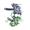
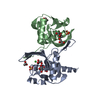
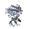
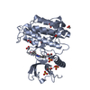
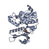
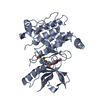
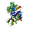
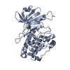
| タイトル | Crystal Structure of a Nudix Protein from Pyrobaculum aerophilum Reveals a Dimer with Intertwined Beta Sheets | ||||||
 要素 要素 | Nudix homolog | ||||||
 キーワード キーワード | STRUCTURAL GENOMICS / UNKNOWN FUNCTION / Nudix/mutT-like fold / mixed alpha/beta / tetramerization due to Hix6x tag / putative Nudix hydrolase | ||||||
| 機能・相同性 |  機能・相同性情報 機能・相同性情報 | ||||||
| 生物種 |   Pyrobaculum aerophilum (古細菌) Pyrobaculum aerophilum (古細菌) | ||||||
| 手法 |  X線回折 / 分子置換 / 解像度: 2.4 Å X線回折 / 分子置換 / 解像度: 2.4 Å | ||||||
 データ登録者 データ登録者 | Wang, S. / Mura, C. / Sawaya, M.R. / Cascio, D. / Eisenberg, D. | ||||||
 引用 引用 | ジャーナル: Acta Crystallogr.,Sect.D / 年: 2002 タイトル: Structure of a Nudix protein from Pyrobaculum aerophilum reveals a dimer with two intersubunit beta-sheets. 著者: Wang, S. / Mura, C. / Sawaya, M.R. / Cascio, D. / Eisenberg, D. | ||||||
| 履歴 |
| ||||||
| Remark 300 | BIOMOLECULE: 1 THIS ENTRY CONTAINS THE CRYSTALLOGRAPHIC ASYMMETRIC UNIT WHICH CONSISTS OF 4 ... BIOMOLECULE: 1 THIS ENTRY CONTAINS THE CRYSTALLOGRAPHIC ASYMMETRIC UNIT WHICH CONSISTS OF 4 CHAIN(S). SEE REMARK 350 FOR INFORMATION ON GENERATING THE BIOLOGICAL MOLECULE(S). The putative biologically-relevant assembly is a dimer, and two of these associate to form a weak tetramer in the asymmetric unit. Chains A+B and C+D form the two dimers. |
- 構造の表示
構造の表示
| 構造ビューア | 分子: MolmilJmol/JSmol |
|---|
- ダウンロードとリンク
-ダウンロード
| PDBx/mmCIF形式 | 1jrk.cif.gz | 129 KB | 表示 | PDBx/mmCIF形式 |
|---|---|---|---|---|
| PDB形式 | pdb1jrk.ent.gz | 104.2 KB | 表示 | PDB形式 |
| PDBx/mmJSON形式 | 1jrk.json.gz | ツリー表示 | PDBx/mmJSON形式 | |
| その他 |  その他のダウンロード その他のダウンロード |
-検証レポート
| アーカイブディレクトリ | https://data.pdbj.org/pub/pdb/validation_reports/jr/1jrkftp://data.pdbj.org/pub/pdb/validation_reports/jr/1jrk | HTTPS FTP |
|---|
-関連構造データ


-リンク
 PDBj
PDBj

- 集合体
集合体
| 登録構造単位 | 
| ||||||||
|---|---|---|---|---|---|---|---|---|---|
| 1 | 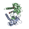
| ||||||||
| 2 | 
| ||||||||
| 3 |
| ||||||||
| 単位格子 |
| ||||||||
| 詳細 | The putative biologically-relevant assembly is a dimer, and two of these associate to form a tetramer in the asymmetric unit. Chains A+B and C+D form the two dimers. |
-要素
| #1: タンパク質 | 分子量: 17813.570 Da / 分子数: 4 / 変異: M16L / 由来タイプ: 組換発現 / 由来: (組換発現) Pyrobaculum aerophilum (古細菌) / プラスミド: PET-22B_MUTT / 生物種 (発現宿主): Escherichia coli / 発現宿主:  #2: 化合物 | ChemComp-MPD / (   分子量: 118.174 Da / 分子数: 6 / 由来タイプ: 合成 / 式: C6H14O2 / コメント: 沈殿剤*YM 分子量: 118.174 Da / 分子数: 6 / 由来タイプ: 合成 / 式: C6H14O2 / コメント: 沈殿剤*YM#3: 水 | ChemComp-HOH / |  分子量: 18.015 Da / 分子数: 173 / 由来タイプ: 天然 / 式: H2O 分子量: 18.015 Da / 分子数: 173 / 由来タイプ: 天然 / 式: H2O |
|---|
-実験情報
-実験
| 実験 | 手法: X線回折 / 使用した結晶の数: 1 |
|---|
- 試料調製
試料調製
| 結晶 | マシュー密度: 2.18 Å3/Da / 溶媒含有率: 43.63 % | ||||||||||||||||||||||||||||||||||||
|---|---|---|---|---|---|---|---|---|---|---|---|---|---|---|---|---|---|---|---|---|---|---|---|---|---|---|---|---|---|---|---|---|---|---|---|---|---|
| 結晶化 | 温度: 293 K / 手法: 蒸気拡散法, ハンギングドロップ法 / pH: 6.2 詳細: Tris, MPD, MES buffer, pH 6.2, VAPOR DIFFUSION, HANGING DROP, temperature 293K | ||||||||||||||||||||||||||||||||||||
| 結晶化 | *PLUS pH: 8 | ||||||||||||||||||||||||||||||||||||
| 溶液の組成 | *PLUS
|
-データ収集
| 回折 | 平均測定温度: 110 K |
|---|---|
| 放射光源 | 由来: 回転陽極 / タイプ: RIGAKU FR-D / 波長: 1.5418 Å |
| 検出器 | タイプ: RIGAKU RAXIS IV++ / 検出器: IMAGE PLATE / 日付: 2001年2月21日 / 詳細: mirrors |
| 放射 | モノクロメーター: Ni filter / プロトコル: SINGLE WAVELENGTH / 単色(M)・ラウエ(L): M / 散乱光タイプ: x-ray |
| 放射波長 | 波長: 1.5418 Å / 相対比: 1 |
| 反射 | 解像度: 2.4→90 Å / Num. all: 23707 / Num. obs: 23707 / % possible obs: 98.2 % / Observed criterion σ(F): -3 / Observed criterion σ(I): -3 / 冗長度: 3.5 % / Rmerge(I) obs: 0.093 / Net I/σ(I): 14.1 |
| 反射 シェル | 解像度: 2.4→2.49 Å / Rmerge(I) obs: 0.465 / Mean I/σ(I) obs: 3 / Num. unique all: 2324 / % possible all: 97 |
| 反射 | *PLUS 最低解像度: 90 Å / Num. measured all: 84048 / Rmerge(I) obs: 0.093 |
| 反射 シェル | *PLUS % possible obs: 97 % / Rmerge(I) obs: 0.465 |
- 解析
解析
| ソフトウェア |
| |||||||||||||||||||||||||
|---|---|---|---|---|---|---|---|---|---|---|---|---|---|---|---|---|---|---|---|---|---|---|---|---|---|---|
| 精密化 | 構造決定の手法: 分子置換 開始モデル: P. aerophilum mutT dimer solved in another space group (P21) by Wang et al. 解像度: 2.4→20 Å Isotropic thermal model: restrained isotropic temperature factors 交差検証法: THROUGHOUT / σ(F): 0 / σ(I): 0 / 立体化学のターゲット値: Engh & Huber
| |||||||||||||||||||||||||
| 原子変位パラメータ | Biso mean: 33.4 Å2 | |||||||||||||||||||||||||
| Refine analyze |
| |||||||||||||||||||||||||
| 精密化ステップ | サイクル: LAST / 解像度: 2.4→20 Å
| |||||||||||||||||||||||||
| 拘束条件 |
| |||||||||||||||||||||||||
| LS精密化 シェル | 解像度: 2.4→2.55 Å / Rfactor Rfree error: 0.027
| |||||||||||||||||||||||||
| 精密化 | *PLUS 最低解像度: 20 Å / % reflection Rfree: 8 % / Rfactor obs: 0.19 / Rfactor Rfree: 0.274 / Rfactor Rwork: 0.19 | |||||||||||||||||||||||||
| 溶媒の処理 | *PLUS | |||||||||||||||||||||||||
| 原子変位パラメータ | *PLUS | |||||||||||||||||||||||||
| 拘束条件 | *PLUS
| |||||||||||||||||||||||||
| LS精密化 シェル | *PLUS Rfactor Rfree: 0.346 / Rfactor Rwork: 0.247 |
