 Movie
Movie Controller


+ Open data

- Basic information
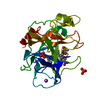
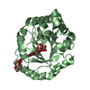
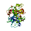
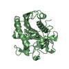
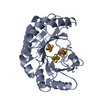
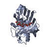
| Entry | Database: PDB / ID: 1jra | ||||||
|---|---|---|---|---|---|---|---|
| Title | Crystal Structure of Erv2p | ||||||
 Components Components | ERV2 PROTEIN, MITOCHONDRIAL | ||||||
 Keywords Keywords | OXIDOREDUCTASE / FAD / sulfhydryl oxidase / helical bundle / CXXC | ||||||
| Function / homology |  Function and homology information Function and homology informationflavin-dependent sulfhydryl oxidase activity / regulation of protein folding in endoplasmic reticulum / thiol oxidase activity / thiol oxidase / fungal-type vacuole / fungal-type vacuole membrane / flavin adenine dinucleotide binding / endoplasmic reticulum membrane / endoplasmic reticulum Similarity search - Function | ||||||
| Biological species |  | ||||||
| Method |  X-RAY DIFFRACTION / MOLECULAR REPLACEMENT / Resolution: 2 Å X-RAY DIFFRACTION / MOLECULAR REPLACEMENT / Resolution: 2 Å | ||||||
 Authors Authors | Gross, E. / Sevier, C.S. / Vala, A. / Kaiser, C.A. / Fass, D. | ||||||
 Citation Citation | Journal: Nat.Struct.Biol. / Year: 2002 Title: A new FAD-binding fold and intersubunit disulfide shuttle in the thiol oxidase Erv2p. Authors: Gross, E. / Sevier, C.S. / Vala, A. / Kaiser, C.A. / Fass, D. | ||||||
| History |
|
- Structure visualization
| Structure viewer | Molecule: MolmilJmol/JSmol |
|---|
- Downloads & links
-Download
| PDBx/mmCIF format | 1jra.cif.gz | 108.1 KB | Display | PDBx/mmCIF format |
|---|---|---|---|---|
| PDB format | pdb1jra.ent.gz | 84 KB | Display | PDB format |
| PDBx/mmJSON format | 1jra.json.gz | Tree view | PDBx/mmJSON format | |
| Others |  Other downloads Other downloads |
-Validation report
| Arichive directory | https://data.pdbj.org/pub/pdb/validation_reports/jr/1jraftp://data.pdbj.org/pub/pdb/validation_reports/jr/1jra | HTTPS FTP |
|---|
-Related structure data
| Related structure data |  1jr8SC S: Starting model for refinement C: citing same article ( |
|---|---|
| Similar structure data |


-Links
 PDBj
PDBj- Assembly
| Deposited unit | 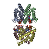
| ||||||||
|---|---|---|---|---|---|---|---|---|---|
| 1 | 
| ||||||||
| 2 | 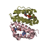
| ||||||||
| Unit cell |
| ||||||||
| Details | The biological assembly is a dimer. The asymmetric unit contains two dimers. |
-Components
| #1: Protein | Mass: 13524.354 Da / Num. of mol.: 4 / Fragment: protease-resistant domain Source method: isolated from a genetically manipulated source Source: (gene. exp.) Gene: ERV2 / Plasmid: pAED4 / Production host:  #2: Chemical | ChemComp-FAD /   Mass: 785.550 Da / Num. of mol.: 4 / Source method: obtained synthetically / Formula: C27H33N9O15P2 / Comment: FAD*YM Mass: 785.550 Da / Num. of mol.: 4 / Source method: obtained synthetically / Formula: C27H33N9O15P2 / Comment: FAD*YM#3: Water | ChemComp-HOH / |  Mass: 18.015 Da / Num. of mol.: 288 / Source method: isolated from a natural source / Formula: H2O Mass: 18.015 Da / Num. of mol.: 288 / Source method: isolated from a natural source / Formula: H2OHas protein modification | Y | |
|---|
-Experimental details
-Experiment
| Experiment | Method: X-RAY DIFFRACTION / Number of used crystals: 1 |
|---|
- Sample preparation
| Crystal | Density Matthews: 2 Å3/Da / Density % sol: 38.44 % | ||||||||||||||||||||||||||||
|---|---|---|---|---|---|---|---|---|---|---|---|---|---|---|---|---|---|---|---|---|---|---|---|---|---|---|---|---|---|
| Crystal grow | Temperature: 293 K / Method: vapor diffusion, hanging drop / pH: 6.2 Details: PEG 8000, sodium cacodylate, magnesium chloride, pH 6.2, VAPOR DIFFUSION, HANGING DROP, temperature 293K | ||||||||||||||||||||||||||||
| Crystal grow | *PLUS | ||||||||||||||||||||||||||||
| Components of the solutions | *PLUS
|
-Data collection
| Diffraction | Mean temperature: 120 K |
|---|---|
| Diffraction source | Source: ROTATING ANODE / Type: RIGAKU RU300 / Wavelength: 1.5418 Å |
| Detector | Type: RIGAKU RAXIS IV / Detector: IMAGE PLATE / Date: Nov 21, 2000 / Details: osmic mirrors |
| Radiation | Monochromator: Ni / Protocol: SINGLE WAVELENGTH / Monochromatic (M) / Laue (L): M / Scattering type: x-ray |
| Radiation wavelength | Wavelength: 1.5418 Å / Relative weight: 1 |
| Reflection | Resolution: 2→20 Å / Num. all: 28911 / Num. obs: 28807 / % possible obs: 99.7 % / Observed criterion σ(F): 0 / Observed criterion σ(I): 0 / Redundancy: 5.3 % / Biso Wilson estimate: 20.9 Å2 / Rmerge(I) obs: 0.082 / Net I/σ(I): 15.1 |
| Reflection shell | Resolution: 2→2.07 Å / Redundancy: 4.8 % / Rmerge(I) obs: 0.461 / Mean I/σ(I) obs: 4.5 / Num. unique all: 2869 / % possible all: 98.3 |
| Reflection | *PLUS % possible obs: 99.8 % |
| Reflection shell | *PLUS % possible obs: 98.3 % |
- Processing
| Software |
| |||||||||||||||||||||||||
|---|---|---|---|---|---|---|---|---|---|---|---|---|---|---|---|---|---|---|---|---|---|---|---|---|---|---|
| Refinement | Method to determine structure: MOLECULAR REPLACEMENT Starting model: PDB entry 1JR8 prior to refinement Resolution: 2→20 Å / Cross valid method: THROUGHOUT / σ(F): 0 / Stereochemistry target values: Engh & Huber
| |||||||||||||||||||||||||
| Refinement step | Cycle: LAST / Resolution: 2→20 Å
| |||||||||||||||||||||||||
| Refine LS restraints |
| |||||||||||||||||||||||||
| Software | *PLUS Name: CNS / Classification: refinement | |||||||||||||||||||||||||
| Refinement | *PLUS Highest resolution: 2 Å / Lowest resolution: 20 Å / Num. reflection obs: 26812 / σ(F): 0 / % reflection Rfree: 7 % / Rfactor obs: 0.196 | |||||||||||||||||||||||||
| Solvent computation | *PLUS | |||||||||||||||||||||||||
| Displacement parameters | *PLUS |
