 Movie
Movie Controller

[English] 日本語
 Yorodumi
- PDB-1jr9: Crystal Structure of manganese superoxide dismutases from Bacillu... -
+ Open data

- Basic information
| Entry | Database: PDB / ID: 1jr9 | ||||||
|---|---|---|---|---|---|---|---|
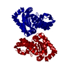
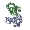
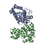
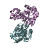
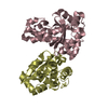
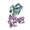
| Title | Crystal Structure of manganese superoxide dismutases from Bacillus halodenitrificans | ||||||
 Components Components | manganese superoxide dismutase | ||||||
 Keywords Keywords | OXIDOREDUCTASE / manganese superoxide dismutase | ||||||
| Function / homology |  Function and homology information Function and homology informationsuperoxide dismutase / superoxide dismutase activity / metal ion binding / cytoplasm Similarity search - Function | ||||||
| Biological species |  Virgibacillus halodenitrificans (bacteria) Virgibacillus halodenitrificans (bacteria) | ||||||
| Method |  X-RAY DIFFRACTION / MOLECULAR REPLACEMENT / Resolution: 2.8 Å X-RAY DIFFRACTION / MOLECULAR REPLACEMENT / Resolution: 2.8 Å | ||||||
 Authors Authors | Liao, J. / Liu, M.Y. / Chang, T. / Li, M. / LeGall, J. / Gui, L.L. / Zhang, J.P. / Jiang, T. / Liang, D.C. / Chang, W.R. | ||||||
 Citation Citation | Journal: J.Struct.Biol. / Year: 2002 Title: Three-dimensional structure of manganese superoxide dismutase from Bacillus halodenitrificans, a component of the so-called "green protein". Authors: Liao, J. / Liu, M.Y. / Chang, T. / Li, M. / Le Gall, J. / Gui, L.L. / Zhang, J.P. / Jiang, T. / Liang, D.C. / Chang, W.R. | ||||||
| History |
|
- Structure visualization
| Structure viewer | Molecule: MolmilJmol/JSmol |
|---|
- Downloads & links
-Download
| PDBx/mmCIF format | 1jr9.cif.gz | 53.9 KB | Display | PDBx/mmCIF format |
|---|---|---|---|---|
| PDB format | pdb1jr9.ent.gz | 38.5 KB | Display | PDB format |
| PDBx/mmJSON format | 1jr9.json.gz | Tree view | PDBx/mmJSON format | |
| Others |  Other downloads Other downloads |
-Validation report
| Arichive directory | https://data.pdbj.org/pub/pdb/validation_reports/jr/1jr9ftp://data.pdbj.org/pub/pdb/validation_reports/jr/1jr9 | HTTPS FTP |
|---|
-Related structure data
| Related structure data |  3mdsS S: Starting model for refinement |
|---|---|
| Similar structure data |


-Links
 PDBj
PDBj
- Assembly
| Deposited unit | 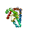
| ||||||||
|---|---|---|---|---|---|---|---|---|---|
| 1 | 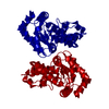
| ||||||||
| Unit cell |
|
-Components
| #1: Protein | Mass: 22688.266 Da / Num. of mol.: 1 / Fragment: green protein / Source method: isolated from a natural source / Details: cytoplasm Source: (natural) Virgibacillus halodenitrificans (bacteria)References: UniProt: Q7SIC3, superoxide dismutase |
|---|---|
| #2: Chemical | ChemComp-MN /   Mass: 54.938 Da / Num. of mol.: 1 / Source method: obtained synthetically / Formula: Mn Mass: 54.938 Da / Num. of mol.: 1 / Source method: obtained synthetically / Formula: Mn |
| #3: Chemical | ChemComp-ZN /   Mass: 65.409 Da / Num. of mol.: 1 / Source method: obtained synthetically / Formula: Zn Mass: 65.409 Da / Num. of mol.: 1 / Source method: obtained synthetically / Formula: Zn |
| #4: Water | ChemComp-HOH /  Mass: 18.015 Da / Num. of mol.: 63 / Source method: isolated from a natural source / Formula: H2O Mass: 18.015 Da / Num. of mol.: 63 / Source method: isolated from a natural source / Formula: H2O |
| Sequence details | A sequence database reference for this protein does not currently exist. |
-Experimental details
-Experiment
| Experiment | Method: X-RAY DIFFRACTION / Number of used crystals: 1 |
|---|
- Sample preparation
| Crystal | Density Matthews: 3.03 Å3/Da / Density % sol: 59.45 % | ||||||||||||||||||||||||||||||||||||
|---|---|---|---|---|---|---|---|---|---|---|---|---|---|---|---|---|---|---|---|---|---|---|---|---|---|---|---|---|---|---|---|---|---|---|---|---|---|
| Crystal grow | Temperature: 281 K / Method: vapor diffusion, hanging drop / pH: 6.5 Details: PEG4000, Sodium Cacodylate, Zinc acetate, pH 6.5, VAPOR DIFFUSION, HANGING DROP, temperature 281K | ||||||||||||||||||||||||||||||||||||
| Crystal | *PLUS Density % sol: 59 % | ||||||||||||||||||||||||||||||||||||
| Crystal grow | *PLUS | ||||||||||||||||||||||||||||||||||||
| Components of the solutions | *PLUS
|
-Data collection
| Diffraction | Mean temperature: 298 K |
|---|---|
| Diffraction source | Source: SEALED TUBE / Type: OTHER / Wavelength: 1.5418 |
| Detector | Type: MARRESEARCH / Detector: IMAGE PLATE / Date: Mar 3, 1999 |
| Radiation | Monochromator: GRAPHITE / Protocol: SINGLE WAVELENGTH / Monochromatic (M) / Laue (L): M / Scattering type: x-ray |
| Radiation wavelength | Wavelength: 1.5418 Å / Relative weight: 1 |
| Reflection | Resolution: 2.8→20 Å / Num. all: 7253 / Num. obs: 5995 / % possible obs: 82.6 % / Observed criterion σ(I): 2 / Redundancy: 18.7 % / Rmerge(I) obs: 0.095 / Net I/σ(I): 19.7 |
| Reflection shell | Resolution: 2.8→2.88 Å / Mean I/σ(I) obs: 3.5 / Num. unique all: 319 / % possible all: 53.8 |
| Reflection | *PLUS Highest resolution: 2.8 Å / Lowest resolution: 20 Å / Num. obs: 4892 / % possible obs: 100 % / Num. measured all: 73544 / Rmerge(I) obs: 0.153 |
| Reflection shell | *PLUS Highest resolution: 3.2 Å / Lowest resolution: 3.3 Å / % possible obs: 100 % / Rmerge(I) obs: 0.527 / Mean I/σ(I) obs: 5.8 |
- Processing
| Software |
| |||||||||||||||||||||||||
|---|---|---|---|---|---|---|---|---|---|---|---|---|---|---|---|---|---|---|---|---|---|---|---|---|---|---|
| Refinement | Method to determine structure: MOLECULAR REPLACEMENT Starting model: PDB ENTRY 3mds Resolution: 2.8→10 Å / Cross valid method: THROUGHOUT / σ(F): 2 / σ(I): 2 / Stereochemistry target values: Engh & Huber
| |||||||||||||||||||||||||
| Refinement step | Cycle: LAST / Resolution: 2.8→10 Å
| |||||||||||||||||||||||||
| Refine LS restraints |
| |||||||||||||||||||||||||
| Refinement | *PLUS Highest resolution: 2.8 Å / Lowest resolution: 10 Å / Rfactor Rwork: 0.22 | |||||||||||||||||||||||||
| Solvent computation | *PLUS | |||||||||||||||||||||||||
| Displacement parameters | *PLUS |
