 Movie
Movie Controller

[English] 日本語
 Yorodumi
- PDB-1jqt: Fitting of L11 protein in the low resolution cryo-EM map of E.col... -
+ Open data

- Basic information
| Entry | Database: PDB / ID: 1jqt | ||||||
|---|---|---|---|---|---|---|---|
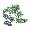
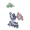
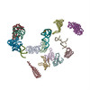
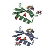
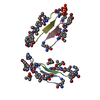
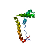
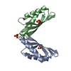
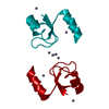
| Title | Fitting of L11 protein in the low resolution cryo-EM map of E.coli 70S ribosome | ||||||
 Components Components | 50S Ribosomal protein L11 | ||||||
 Keywords Keywords | RIBOSOME / L11 / cryo-EM / 70S E.coli ribosome | ||||||
| Function / homology |  Function and homology information Function and homology informationlarge ribosomal subunit rRNA binding / cytosolic large ribosomal subunit / structural constituent of ribosome / translation Similarity search - Function | ||||||
| Biological species |  | ||||||
| Method | ELECTRON MICROSCOPY / single particle reconstruction / Molecular Modeling based on crystal structures / cryo EM / Resolution: 18 Å | ||||||
 Authors Authors | Agrawal, R.K. / Linde, J. / Segupta, J. / Nierhaus, K.H. / Frank, J. | ||||||
 Citation Citation | Journal: J Mol Biol / Year: 2001 Title: Localization of L11 protein on the ribosome and elucidation of its involvement in EF-G-dependent translocation. Authors: R K Agrawal / J Linde / J Sengupta / K H Nierhaus / J Frank /  Abstract: L11 protein is located at the base of the L7/L12 stalk of the 50 S subunit of the Escherichia coli ribosome. Because of the flexible nature of the region, recent X-ray crystallographic studies of the ...L11 protein is located at the base of the L7/L12 stalk of the 50 S subunit of the Escherichia coli ribosome. Because of the flexible nature of the region, recent X-ray crystallographic studies of the 50 S subunit failed to locate the N-terminal domain of the protein. We have determined the position of the complete L11 protein by comparing a three-dimensional cryo-EM reconstruction of the 70 S ribosome, isolated from a mutant lacking ribosomal protein L11, with the three-dimensional map of the wild-type ribosome. Fitting of the X-ray coordinates of L11-23 S RNA complex and EF-G into the cryo-EM maps combined with molecular modeling, reveals that, following EF-G-dependent GTP hydrolysis, domain V of EF-G intrudes into the cleft between the 23 S ribosomal RNA and the N-terminal domain of L11 (where the antibiotic thiostrepton binds), causing the N-terminal domain to move and thereby inducing the formation of the arc-like connection with the G' domain of EF-G. The results provide a new insight into the mechanism of EF-G-dependent translocation. #1: Journal: Cell(Cambridge,Mass.) / Year: 1999Title: A Detailed View of a Ribosomal Active Site: The Structure of the L11-RNA Complex Authors: Wimberly, B.T. / Guymon, R. / McCutcheon, J.P. / White, S.W. / Ramakrishnan, V. #2: Journal: Cell(Cambridge,Mass.) / Year: 2000Title: EF-G-dependent GTP hydrolysis induces solution structure of the E. coli 70S ribosome at 11.5A resolution Authors: Gabashvili, I.S. / Agrawal, R.K. / Spahn, C.M.T. / Grassucci, R.A. / Svergun, D.I. / Frank, J. / Penczek, P. #3: Journal: Nat.Struct.Biol. / Year: 1999Title: EF-G-dependent GTP hydrolysis induces translocation accompanied by large conformational changes in the 70S ribosome Authors: Agrawal, R.K. / Heagle, A.B. / Penczek, P. / Grassucci, R.A. / Frank, J. | ||||||
| History |
|
- Structure visualization
| Movie |
Movie viewer |
|---|---|
| Structure viewer | Molecule: MolmilJmol/JSmol |
- Downloads & links
-Download
| PDBx/mmCIF format | 1jqt.cif.gz | 15 KB | Display | PDBx/mmCIF format |
|---|---|---|---|---|
| PDB format | pdb1jqt.ent.gz | 6.2 KB | Display | PDB format |
| PDBx/mmJSON format | 1jqt.json.gz | Tree view | PDBx/mmJSON format | |
| Others |  Other downloads Other downloads |
-Validation report
| Arichive directory | https://data.pdbj.org/pub/pdb/validation_reports/jq/1jqtftp://data.pdbj.org/pub/pdb/validation_reports/jq/1jqt | HTTPS FTP |
|---|
-Related structure data
-Links
 PDBj
PDBj


- Assembly
| Deposited unit | 
|
|---|---|
| 1 |
|
-Components
| #1: Protein | Mass: 14865.637 Da / Num. of mol.: 1 / Source method: isolated from a natural source Details: L11 from E. coli 70S ribosome modeled by crystal structure of L11 from Thermatogoma maritima Source: (natural) |
|---|
-Experimental details
-Experiment
| Experiment | Method: ELECTRON MICROSCOPY |
|---|---|
| EM experiment | Aggregation state: PARTICLE / 3D reconstruction method: single particle reconstruction |
| NMR details | Text: This structure was generated by fitting the X-ray crystal structure of L11 into the 70S E. coli ribosome map. L11 (linker region between N and C terminal) was modeled to accommodate the observed densities. |
- Sample preparation
| Component | Name: 70S ribosome / Type: RIBOSOME |
|---|---|
| Specimen | Embedding applied: NO / Shadowing applied: NO / Staining applied: NO / Vitrification applied: YES |
| Crystal grow | *PLUS Method: electron microscopy |
- Electron microscopy imaging
| Microscopy | Model: FEI/PHILIPS EM420 |
|---|---|
| Electron gun | Electron source: OTHER / Illumination mode: FLOOD BEAM |
| Electron lens | Mode: BRIGHT FIELD / Nominal magnification: 52000 X |
| Specimen holder | Specimen holder model: GATAN LIQUID NITROGEN |
| Image recording | Electron dose: 10 e/Å2 / Film or detector model: GENERIC FILM |
- Processing
| Symmetry | Point symmetry: C1 (asymmetric) | ||||||||||||||||||||
|---|---|---|---|---|---|---|---|---|---|---|---|---|---|---|---|---|---|---|---|---|---|
| 3D reconstruction | Resolution: 18 Å / Resolution method: FSC 0.5 CUT-OFF / Num. of particles: 36113 / Symmetry type: POINT | ||||||||||||||||||||
| Atomic model building | Protocol: OTHER / Space: REAL / Target criteria: VISUAL AGREEMENT Details: REFINEMENT PROTOCOL--MANUAL DETAILS--This structure was generated by fitting the X-ray crystal structure of L11 into the 70S E. coli ribosome map. L11 (linker region between N and C terminal) ...Details: REFINEMENT PROTOCOL--MANUAL DETAILS--This structure was generated by fitting the X-ray crystal structure of L11 into the 70S E. coli ribosome map. L11 (linker region between N and C terminal) was modeled to accommodate the observed densities. Conformational changes occur in protein L11 and EF-G due to the binding of EF-G to the 70S ribosome. These changed conformations were modeled based on the fitting of the crystal coordinates to the low resolution ribosome map (factor-bound) and energy minimizing the fitted structures. | ||||||||||||||||||||
| Refinement step | Cycle: LAST
| ||||||||||||||||||||
| NMR software |
| ||||||||||||||||||||
| Refinement | Method: Molecular Modeling based on crystal structures / Software ordinal: 1 Details: Conformational changes occur in protein L11 when it is bound to 70S ribosome. This changed conformation was modeled based on the fitting of the crystal coordinates to the low resolution ...Details: Conformational changes occur in protein L11 when it is bound to 70S ribosome. This changed conformation was modeled based on the fitting of the crystal coordinates to the low resolution ribosome map and energy minimizing the fitted structures. |
