 Movie
Movie Controller

[English] 日本語
 Yorodumi
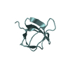
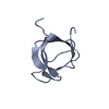
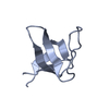
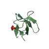
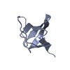
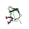
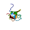
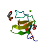
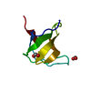
- PDB-1jo8: Structural analysis of the yeast actin binding protein Abp1 SH3 domain -
+ Open data

- Basic information
| Entry | Database: PDB / ID: 1jo8 | ||||||
|---|---|---|---|---|---|---|---|
| Title | Structural analysis of the yeast actin binding protein Abp1 SH3 domain | ||||||
 Components Components | ACTIN BINDING PROTEIN | ||||||
 Keywords Keywords | STRUCTURAL PROTEIN / SH3 domain Actin-binding-protein | ||||||
| Function / homology |  Function and homology information Function and homology informationprotein localization to actin cortical patch / positive regulation of Arp2/3 complex-mediated actin nucleation / site of polarized growth / actin cortical patch assembly / actin cortical patch / regulation of actin filament polymerization / barbed-end actin filament capping / mating projection tip / cortical actin cytoskeleton / actin filament binding ...protein localization to actin cortical patch / positive regulation of Arp2/3 complex-mediated actin nucleation / site of polarized growth / actin cortical patch assembly / actin cortical patch / regulation of actin filament polymerization / barbed-end actin filament capping / mating projection tip / cortical actin cytoskeleton / actin filament binding / cell cortex / cytoplasm Similarity search - Function | ||||||
| Biological species |  | ||||||
| Method |  X-RAY DIFFRACTION / SYNCHROTRON / MOLECULAR REPLACEMENT / Resolution: 1.3 Å X-RAY DIFFRACTION / SYNCHROTRON / MOLECULAR REPLACEMENT / Resolution: 1.3 Å | ||||||
 Authors Authors | Fazi, B. / Cope, M.J. / Douangamath, A. / Ferracuti, S. / Schirwitz, K. / Zucconi, A. / Drubin, D.G. / Wilmanns, M. / Cesareni, G. / Castagnoli, L. | ||||||
 Citation Citation | Journal: J.Biol.Chem. / Year: 2002 Title: Unusual binding properties of the SH3 domain of the yeast actin-binding protein Abp1: structural and functional analysis. Authors: Fazi, B. / Cope, M.J. / Douangamath, A. / Ferracuti, S. / Schirwitz, K. / Zucconi, A. / Drubin, D.G. / Wilmanns, M. / Cesareni, G. / Castagnoli, L. | ||||||
| History |
|
- Structure visualization
| Structure viewer | Molecule: MolmilJmol/JSmol |
|---|
- Downloads & links
-Download
| PDBx/mmCIF format | 1jo8.cif.gz | 41.9 KB | Display | PDBx/mmCIF format |
|---|---|---|---|---|
| PDB format | pdb1jo8.ent.gz | 30.1 KB | Display | PDB format |
| PDBx/mmJSON format | 1jo8.json.gz | Tree view | PDBx/mmJSON format | |
| Others |  Other downloads Other downloads |
-Validation report
| Arichive directory | https://data.pdbj.org/pub/pdb/validation_reports/jo/1jo8ftp://data.pdbj.org/pub/pdb/validation_reports/jo/1jo8 | HTTPS FTP |
|---|
-Related structure data
| Related structure data | 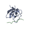 1ckaS S: Starting model for refinement |
|---|---|
| Similar structure data |

-Links
 PDBj
PDBj
- Assembly
| Deposited unit | 
| ||||||||||
|---|---|---|---|---|---|---|---|---|---|---|---|
| 1 |
| ||||||||||
| Unit cell |
|
-Components
| #1: Protein | Mass: 6663.082 Da / Num. of mol.: 1 / Fragment: SH3 domain Source method: isolated from a genetically manipulated source Source: (gene. exp.) Plasmid: pRSETA / Production host:  | ||
|---|---|---|---|
| #2: Chemical |   Mass: 96.063 Da / Num. of mol.: 2 / Source method: obtained synthetically / Formula: SO4 Mass: 96.063 Da / Num. of mol.: 2 / Source method: obtained synthetically / Formula: SO4#3: Water | ChemComp-HOH / |  Mass: 18.015 Da / Num. of mol.: 129 / Source method: isolated from a natural source / Formula: H2O Mass: 18.015 Da / Num. of mol.: 129 / Source method: isolated from a natural source / Formula: H2O |
-Experimental details
-Experiment
| Experiment | Method: X-RAY DIFFRACTION / Number of used crystals: 1 |
|---|
- Sample preparation
| Crystal | Density Matthews: 2.04 Å3/Da / Density % sol: 39.6 % | ||||||||||||||||||||||||
|---|---|---|---|---|---|---|---|---|---|---|---|---|---|---|---|---|---|---|---|---|---|---|---|---|---|
| Crystal grow | Temperature: 298 K / Method: vapor diffusion, sitting drop / pH: 8 Details: Ammonium sulfate, bis-tris-propane, pH 8.0, VAPOR DIFFUSION, SITTING DROP at 298K | ||||||||||||||||||||||||
| Crystal grow | *PLUS | ||||||||||||||||||||||||
| Components of the solutions | *PLUS
|
-Data collection
| Diffraction | Mean temperature: 100 K |
|---|---|
| Diffraction source | Source: SYNCHROTRON / Site: EMBL/DESY, HAMBURG  / Beamline: BW7B / Wavelength: 0.842 Å / Beamline: BW7B / Wavelength: 0.842 Å |
| Detector | Type: MARRESEARCH / Detector: IMAGE PLATE / Date: Sep 15, 2000 |
| Radiation | Monochromator: SAGITALLY FOCUSED Si(111) / Protocol: SINGLE WAVELENGTH / Monochromatic (M) / Laue (L): M / Scattering type: x-ray |
| Radiation wavelength | Wavelength: 0.842 Å / Relative weight: 1 |
| Reflection | Resolution: 1.3→25 Å / Num. all: 57575 / Num. obs: 56424 / % possible obs: 98 % / Redundancy: 4.1 % / Biso Wilson estimate: 9.8 Å2 / Rmerge(I) obs: 0.056 / Net I/σ(I): 20.3 |
| Reflection shell | Resolution: 1.3→1.32 Å / Rmerge(I) obs: 0.266 / Mean I/σ(I) obs: 4 / Num. unique all: 617 / % possible all: 88.6 |
| Reflection | *PLUS Lowest resolution: 40 Å / Num. obs: 13742 / % possible obs: 98 % / Num. measured all: 56424 |
| Reflection shell | *PLUS % possible obs: 88.6 % |
- Processing
| Software |
| ||||||||||||||||||||||||||||||||||||||||||||||||
|---|---|---|---|---|---|---|---|---|---|---|---|---|---|---|---|---|---|---|---|---|---|---|---|---|---|---|---|---|---|---|---|---|---|---|---|---|---|---|---|---|---|---|---|---|---|---|---|---|---|
| Refinement | Method to determine structure: MOLECULAR REPLACEMENT Starting model: PDB entry 1CKA Resolution: 1.3→17.51 Å / Cross valid method: THROUGHOUT / σ(F): 0 / Stereochemistry target values: Engh & Huber
| ||||||||||||||||||||||||||||||||||||||||||||||||
| Displacement parameters | Biso mean: 8.742 Å2
| ||||||||||||||||||||||||||||||||||||||||||||||||
| Refinement step | Cycle: LAST / Resolution: 1.3→17.51 Å
| ||||||||||||||||||||||||||||||||||||||||||||||||
| Refine LS restraints |
| ||||||||||||||||||||||||||||||||||||||||||||||||
| LS refinement shell | Resolution: 1.3→1.334 Å / Total num. of bins used: 20
| ||||||||||||||||||||||||||||||||||||||||||||||||
| Software | *PLUS Name: REFMAC / Version: 5 / Classification: refinement | ||||||||||||||||||||||||||||||||||||||||||||||||
| Refinement | *PLUS σ(F): 0 / % reflection Rfree: 5 % | ||||||||||||||||||||||||||||||||||||||||||||||||
| Solvent computation | *PLUS | ||||||||||||||||||||||||||||||||||||||||||||||||
| Displacement parameters | *PLUS | ||||||||||||||||||||||||||||||||||||||||||||||||
| LS refinement shell | *PLUS Rfactor Rfree: 0.194 / Rfactor Rwork: 0.171 / Total num. of bins used: 20 |
