 Movie
Movie Controller


+ Open data

- Basic information
| Entry | Database: PDB / ID: 1jnd | |||||||||
|---|---|---|---|---|---|---|---|---|---|---|
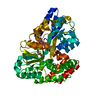
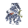
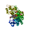
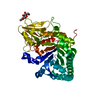
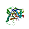
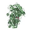
| Title | Crystal structure of imaginal disc growth factor-2 | |||||||||
 Components Components | Imaginal disc growth factor-2 | |||||||||
 Keywords Keywords | HORMONE/GROWTH FACTOR / IDGF / imaginal disc / growth factor / chitinase / insulin receptor / heparin / HORMONE-GROWTH FACTOR COMPLEX | |||||||||
| Function / homology |  Function and homology information Function and homology informationimaginal disc growth factor receptor binding / Digestion of dietary carbohydrate / imaginal disc development / Neutrophil degranulation / chitin catabolic process / chitin binding / carbohydrate metabolic process / positive regulation of cell population proliferation / negative regulation of apoptotic process / : / extracellular region Similarity search - Function | |||||||||
| Biological species |  | |||||||||
| Method |  X-RAY DIFFRACTION / SYNCHROTRON / MOLECULAR REPLACEMENT / Resolution: 1.3 Å X-RAY DIFFRACTION / SYNCHROTRON / MOLECULAR REPLACEMENT / Resolution: 1.3 Å | |||||||||
 Authors Authors | Varela, P.F. / Llera, A.S. / Mariuzza, R.A. / Tormo, J. | |||||||||
 Citation Citation | Journal: J.Biol.Chem. / Year: 2002 Title: Crystal structure of imaginal disc growth factor-2. A member of a new family of growth-promoting glycoproteins from Drosophila melanogaster. Authors: Varela, P.F. / Llera, A.S. / Mariuzza, R.A. / Tormo, J. | |||||||||
| History |
|
- Structure visualization
| Structure viewer | Molecule: MolmilJmol/JSmol |
|---|
- Downloads & links
-Download
| PDBx/mmCIF format | 1jnd.cif.gz | 111.1 KB | Display | PDBx/mmCIF format |
|---|---|---|---|---|
| PDB format | pdb1jnd.ent.gz | 83.3 KB | Display | PDB format |
| PDBx/mmJSON format | 1jnd.json.gz | Tree view | PDBx/mmJSON format | |
| Others |  Other downloads Other downloads |
-Validation report
| Arichive directory | https://data.pdbj.org/pub/pdb/validation_reports/jn/1jndftp://data.pdbj.org/pub/pdb/validation_reports/jn/1jnd | HTTPS FTP |
|---|
-Related structure data
| Related structure data |  1jneC 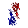 1ctnS S: Starting model for refinement C: citing same article ( |
|---|---|
| Similar structure data |


-Links
 PDBj
PDBj


- Assembly
| Deposited unit | 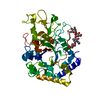
| ||||||||
|---|---|---|---|---|---|---|---|---|---|
| 1 |
| ||||||||
| Unit cell |
| ||||||||
| Components on special symmetry positions |
|
-Components
| #1: Protein | Mass: 46952.762 Da / Num. of mol.: 1 / Source method: isolated from a natural source / Source: (natural) |
|---|---|
| #2: Polysaccharide | alpha-D-mannopyranose-(1-3)-beta-D-mannopyranose-(1-4)-2-acetamido-2-deoxy-beta-D-glucopyranose-(1- ...alpha-D-mannopyranose-(1-3)-beta-D-mannopyranose-(1-4)-2-acetamido-2-deoxy-beta-D-glucopyranose-(1-4)-2-acetamido-2-deoxy-beta-D-glucopyranose Source method: isolated from a genetically manipulated source |
| #3: Water | ChemComp-HOH /  Mass: 18.015 Da / Num. of mol.: 700 / Source method: isolated from a natural source / Formula: H2O Mass: 18.015 Da / Num. of mol.: 700 / Source method: isolated from a natural source / Formula: H2O |
| Has protein modification | Y |
-Experimental details
-Experiment
| Experiment | Method: X-RAY DIFFRACTION / Number of used crystals: 1 |
|---|
- Sample preparation
| Crystal | Density Matthews: 3.13 Å3/Da / Density % sol: 60.66 % | ||||||||||||||||||||||||
|---|---|---|---|---|---|---|---|---|---|---|---|---|---|---|---|---|---|---|---|---|---|---|---|---|---|
| Crystal grow | Temperature: 277 K / Method: vapor diffusion, hanging drop / pH: 5 Details: PEG 4000, sodium acetate, pH 5.0, VAPOR DIFFUSION, HANGING DROP, temperature 277K | ||||||||||||||||||||||||
| Crystal grow | *PLUS Temperature: 4 ℃ / Method: unknown | ||||||||||||||||||||||||
| Components of the solutions | *PLUS
|
-Data collection
| Diffraction | Mean temperature: 100 K |
|---|---|
| Diffraction source | Source: SYNCHROTRON / Site: NSLS  / Beamline: X12B / Wavelength: 0.9464 / Wavelength: 0.9464 Å / Beamline: X12B / Wavelength: 0.9464 / Wavelength: 0.9464 Å |
| Detector | Type: ADSC QUANTUM 4 / Detector: CCD / Date: Nov 5, 1999 |
| Radiation | Protocol: SINGLE WAVELENGTH / Monochromatic (M) / Laue (L): M / Scattering type: x-ray |
| Radiation wavelength | Wavelength: 0.9464 Å / Relative weight: 1 |
| Reflection | Resolution: 1.3→100 Å / Num. obs: 129904 / % possible obs: 90.3 % / Redundancy: 1.1 % / Rsym value: 0.023 |
| Reflection shell | Resolution: 1.3→1.33 Å / Redundancy: 1.76 % / Mean I/σ(I) obs: 2.88 / Rsym value: 0.257 / % possible all: 53.5 |
| Reflection | *PLUS Lowest resolution: 100 Å / Num. measured all: 758311 / Rmerge(I) obs: 0.023 |
| Reflection shell | *PLUS % possible obs: 53.5 % / Rmerge(I) obs: 0.257 |
- Processing
| Software |
| ||||||||||||||||||||
|---|---|---|---|---|---|---|---|---|---|---|---|---|---|---|---|---|---|---|---|---|---|
| Refinement | Method to determine structure: MOLECULAR REPLACEMENT Starting model: PDB ENTRY 1CTN Resolution: 1.3→100 Å / Isotropic thermal model: Isotropic / Stereochemistry target values: Engh & Huber
| ||||||||||||||||||||
| Refinement step | Cycle: LAST / Resolution: 1.3→100 Å
| ||||||||||||||||||||
| Refine LS restraints |
| ||||||||||||||||||||
| Software | *PLUS Name: SHELXL / Version: 97 / Classification: refinement | ||||||||||||||||||||
| Refinement | *PLUS Lowest resolution: 100 Å / % reflection Rfree: 1.8 % | ||||||||||||||||||||
| Solvent computation | *PLUS | ||||||||||||||||||||
| Displacement parameters | *PLUS | ||||||||||||||||||||
| Refine LS restraints | *PLUS
|
