 Movie
Movie Controller

[English] 日本語
 Yorodumi
- PDB-1jms: Crystal Structure of the Catalytic Core of Murine Terminal Deoxyn... -
+ Open data

- Basic information
| Entry | Database: PDB / ID: 1jms | ||||||
|---|---|---|---|---|---|---|---|
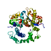
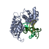
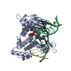
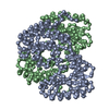
| Title | Crystal Structure of the Catalytic Core of Murine Terminal Deoxynucleotidyl Transferase | ||||||
 Components Components | TERMINAL DEOXYNUCLEOTIDYLTRANSFERASE | ||||||
 Keywords Keywords | TRANSFERASE / polymerase / nucleotidyl transferase | ||||||
| Function / homology |  Function and homology information Function and homology informationDNA nucleotidylexotransferase / DNA nucleotidylexotransferase activity / DNA modification / Hydrolases; Acting on ester bonds; Exodeoxyribonucleases producing 5'-phosphomonoesters / DNA metabolic process / response to ATP / euchromatin / double-strand break repair via nonhomologous end joining / nuclear matrix / DNA-directed DNA polymerase activity ...DNA nucleotidylexotransferase / DNA nucleotidylexotransferase activity / DNA modification / Hydrolases; Acting on ester bonds; Exodeoxyribonucleases producing 5'-phosphomonoesters / DNA metabolic process / response to ATP / euchromatin / double-strand break repair via nonhomologous end joining / nuclear matrix / DNA-directed DNA polymerase activity / hydrolase activity / chromatin / DNA binding / nucleoplasm / metal ion binding / nucleus / cytosol Similarity search - Function | ||||||
| Biological species |  | ||||||
| Method |  X-RAY DIFFRACTION / SYNCHROTRON / MIR / Resolution: 2.36 Å X-RAY DIFFRACTION / SYNCHROTRON / MIR / Resolution: 2.36 Å | ||||||
 Authors Authors | Delarue, M. / Boule, J.B. / Lescar, J. / Expert-Bezancon, N. / Sukumar, N. / Jourdan, N. / Rougeon, F. / Papanicolaou, C. | ||||||
 Citation Citation | Journal: Embo J. / Year: 2002 Title: Crystal structures of a template-independent DNA polymerase: murine terminal deoxynucleotidyltransferase. Authors: Delarue, M. / Boule, J.B. / Lescar, J. / Expert-Bezancon, N. / Jourdan, N. / Sukumar, N. / Rougeon, F. / Papanicolaou, C. #1: Journal: Acta Crystallogr.,Sect.D / Year: 2000Title: Crystallization of the Catalytic Domain of Murine Deoxynucleotidyl Transferase Authors: Sukumar, N. / Boule, J.B. / Expert-Bezancon, N. / Jourdan, N. / Lescar, J. / Rougeon, F. / Papanicolaou, C. / Delarue, M. | ||||||
| History |
|
- Structure visualization
| Structure viewer | Molecule: MolmilJmol/JSmol |
|---|
- Downloads & links
-Download
| PDBx/mmCIF format | 1jms.cif.gz | 90.2 KB | Display | PDBx/mmCIF format |
|---|---|---|---|---|
| PDB format | pdb1jms.ent.gz | 67.7 KB | Display | PDB format |
| PDBx/mmJSON format | 1jms.json.gz | Tree view | PDBx/mmJSON format | |
| Others |  Other downloads Other downloads |
-Validation report
| Arichive directory | https://data.pdbj.org/pub/pdb/validation_reports/jm/1jmsftp://data.pdbj.org/pub/pdb/validation_reports/jm/1jms | HTTPS FTP |
|---|
-Related structure data


-Links
 PDBj
PDBj


- Assembly
| Deposited unit | 
| ||||||||
|---|---|---|---|---|---|---|---|---|---|
| 1 |
| ||||||||
| Unit cell |
|
-Components
| #1: Protein | Mass: 43602.648 Da / Num. of mol.: 1 Source method: isolated from a genetically manipulated source Details: short isoform / Source: (gene. exp.)  |
|---|---|
| #2: Chemical | ChemComp-MG /   Mass: 24.305 Da / Num. of mol.: 1 / Source method: obtained synthetically / Formula: Mg Mass: 24.305 Da / Num. of mol.: 1 / Source method: obtained synthetically / Formula: Mg |
| #3: Chemical | ChemComp-NA /   Mass: 22.990 Da / Num. of mol.: 1 / Source method: obtained synthetically / Formula: Na Mass: 22.990 Da / Num. of mol.: 1 / Source method: obtained synthetically / Formula: Na |
| #4: Water | ChemComp-HOH /  Mass: 18.015 Da / Num. of mol.: 214 / Source method: isolated from a natural source / Formula: H2O Mass: 18.015 Da / Num. of mol.: 214 / Source method: isolated from a natural source / Formula: H2O |
-Experimental details
-Experiment
| Experiment | Method: X-RAY DIFFRACTION / Number of used crystals: 1 |
|---|
- Sample preparation
| Crystal | Density Matthews: 2.55 Å3/Da / Density % sol: 50 % | ||||||||||||||||||||||||||||||||||||||||||
|---|---|---|---|---|---|---|---|---|---|---|---|---|---|---|---|---|---|---|---|---|---|---|---|---|---|---|---|---|---|---|---|---|---|---|---|---|---|---|---|---|---|---|---|
| Crystal grow | Temperature: 290 K / Method: vapor diffusion, hanging drop / pH: 6 Details: PEG 4000, LiCl 1M, pH 6.0, VAPOR DIFFUSION, HANGING DROP, temperature 290K | ||||||||||||||||||||||||||||||||||||||||||
| Crystal grow | *PLUS Temperature: 253 KDetails: Sukumar, N., (2000) Acta Crystallogr., Sect.D, 56, 1662. pH: 6.8 | ||||||||||||||||||||||||||||||||||||||||||
| Components of the solutions | *PLUS
|
-Data collection
| Diffraction | Mean temperature: 110 K |
|---|---|
| Diffraction source | Source: SYNCHROTRON / Site: ESRF  / Beamline: ID14-3 / Wavelength: 0.94 Å / Beamline: ID14-3 / Wavelength: 0.94 Å |
| Detector | Type: MARRESEARCH / Detector: CCD / Date: Apr 25, 1999 |
| Radiation | Monochromator: diamond / Protocol: SINGLE WAVELENGTH / Monochromatic (M) / Laue (L): M / Scattering type: x-ray |
| Radiation wavelength | Wavelength: 0.94 Å / Relative weight: 1 |
| Reflection | Resolution: 2.35→25 Å / Num. all: 19351 / Num. obs: 19351 / % possible obs: 99.8 % / Observed criterion σ(F): 0 / Observed criterion σ(I): 0 / Redundancy: 5.2 % / Biso Wilson estimate: 46.3 Å2 / Rmerge(I) obs: 0.059 / Rsym value: 0.059 / Net I/σ(I): 25.3 |
| Reflection shell | Resolution: 2.35→2.4 Å / Redundancy: 4.1 % / Rmerge(I) obs: 0.34 / Mean I/σ(I) obs: 4.1 / Rsym value: 0.34 / % possible all: 98.4 |
| Reflection | *PLUS Lowest resolution: 20 Å / Num. measured all: 99454 |
| Reflection shell | *PLUS % possible obs: 98.4 % / Num. unique obs: 1224 / Rmerge(I) obs: 0.34 |
- Processing
| Software |
| ||||||||||||||||||||||||||||
|---|---|---|---|---|---|---|---|---|---|---|---|---|---|---|---|---|---|---|---|---|---|---|---|---|---|---|---|---|---|
| Refinement | Method to determine structure: MIR / Resolution: 2.36→18 Å / Data cutoff high rms absF: 1000 / Isotropic thermal model: yes / Cross valid method: THROUGHOUT / σ(F): 0 / Stereochemistry target values: MLF
| ||||||||||||||||||||||||||||
| Solvent computation | Solvent model: FLAT MODEL / Bsol: 38.91 Å2 / ksol: 0.3511 e/Å3 | ||||||||||||||||||||||||||||
| Displacement parameters | Biso mean: 39.2 Å2
| ||||||||||||||||||||||||||||
| Refinement step | Cycle: LAST / Resolution: 2.36→18 Å
| ||||||||||||||||||||||||||||
| Refine LS restraints |
| ||||||||||||||||||||||||||||
| LS refinement shell | Resolution: 2.36→2.4 Å / Total num. of bins used: 19
| ||||||||||||||||||||||||||||
| Xplor file |
| ||||||||||||||||||||||||||||
| Software | *PLUS Name: CNS / Classification: refinement | ||||||||||||||||||||||||||||
| Refinement | *PLUS Highest resolution: 2.35 Å / Lowest resolution: 18 Å / Num. reflection obs: 17984 / σ(F): 0 / Num. reflection Rfree: 973 / % reflection Rfree: 5 % / Rfactor obs: 0.21 / Rfactor Rfree: 0.258 | ||||||||||||||||||||||||||||
| Solvent computation | *PLUS | ||||||||||||||||||||||||||||
| Displacement parameters | *PLUS Biso mean: 39.2 Å2 | ||||||||||||||||||||||||||||
| Refine LS restraints | *PLUS
| ||||||||||||||||||||||||||||
| LS refinement shell | *PLUS Rfactor Rfree: 0.38 / % reflection Rfree: 5 % |
