 Movie
Movie Controller

[English] 日本語
 Yorodumi
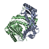
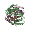
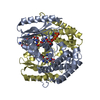
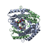
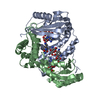
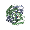
- PDB-1kan: MOLECULAR STRUCTURE OF KANAMYCIN NUCLEOTIDYLTRANSFERASE DETERMINE... -
+ Open data

- Basic information
| Entry | Database: PDB / ID: 1kan | ||||||
|---|---|---|---|---|---|---|---|
| Title | MOLECULAR STRUCTURE OF KANAMYCIN NUCLEOTIDYLTRANSFERASE DETERMINED TO 3.0-ANGSTROMS RESOLUTION | ||||||
 Components Components | KANAMYCIN NUCLEOTIDYLTRANSFERASE | ||||||
 Keywords Keywords | NUCLEOTIDYLTRANSFERASE | ||||||
| Function / homology | Kanamycin nucleotidyltransferase, C-terminal / KNTase C-terminal domain / nucleotidyltransferase activity / Nucleotidyltransferase superfamily / Transferases; Transferring phosphorus-containing groups; Nucleotidyltransferases / response to antibiotic / ATP binding / Aminoglycoside nucleotidyltransferase (4') Function and homology information Function and homology information | ||||||
| Biological species |   Staphylococcus aureus (bacteria) Staphylococcus aureus (bacteria) | ||||||
| Method |  X-RAY DIFFRACTION / Resolution: 3 Å X-RAY DIFFRACTION / Resolution: 3 Å | ||||||
 Authors Authors | Holden, H.M. / Rayment, I. / Sakon, J. | ||||||
 Citation Citation | Journal: Biochemistry / Year: 1993 Title: Molecular structure of kanamycin nucleotidyltransferase determined to 3.0-A resolution. Authors: Sakon, J. / Liao, H.H. / Kanikula, A.M. / Benning, M.M. / Rayment, I. / Holden, H.M. | ||||||
| History |
|
- Structure visualization
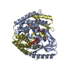
| Structure viewer | Molecule: MolmilJmol/JSmol |
|---|
- Downloads & links
-Download
| PDBx/mmCIF format | 1kan.cif.gz | 27 KB | Display | PDBx/mmCIF format |
|---|---|---|---|---|
| PDB format | pdb1kan.ent.gz | 14.5 KB | Display | PDB format |
| PDBx/mmJSON format | 1kan.json.gz | Tree view | PDBx/mmJSON format | |
| Others |  Other downloads Other downloads |
-Validation report
| Arichive directory | https://data.pdbj.org/pub/pdb/validation_reports/ka/1kanftp://data.pdbj.org/pub/pdb/validation_reports/ka/1kan | HTTPS FTP |
|---|
-Related structure data
| Similar structure data |
|---|

-Links
 PDBj
PDBj
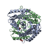
- Assembly
| Deposited unit | 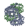
| ||||||||
|---|---|---|---|---|---|---|---|---|---|
| 1 |
| ||||||||
| Unit cell |
|
-Components
| #1: Protein | Mass: 28901.689 Da / Num. of mol.: 2 Source method: isolated from a genetically manipulated source Source: (gene. exp.) Staphylococcus aureus (bacteria)References: UniProt: P05057, Transferases; Transferring phosphorus-containing groups; Nucleotidyltransferases |
|---|
-Experimental details
-Experiment
| Experiment | Method: X-RAY DIFFRACTION |
|---|
- Sample preparation
| Crystal | Density Matthews: 2.95 Å3/Da / Density % sol: 58.31 % | ||||||||||||||||||||||||||||||||||||||||||||||||
|---|---|---|---|---|---|---|---|---|---|---|---|---|---|---|---|---|---|---|---|---|---|---|---|---|---|---|---|---|---|---|---|---|---|---|---|---|---|---|---|---|---|---|---|---|---|---|---|---|---|
| Crystal grow | *PLUS Temperature: 4 ℃ / pH: 8 / Method: vapor diffusion, hanging drop | ||||||||||||||||||||||||||||||||||||||||||||||||
| Components of the solutions | *PLUS
|
-Data collection
| Radiation | Scattering type: x-ray |
|---|---|
| Radiation wavelength | Relative weight: 1 |
| Reflection | *PLUS Highest resolution: 3 Å / Lowest resolution: 9999 Å / Num. obs: 13914 / % possible obs: 95 % / Num. measured all: 70244 / Rmerge(I) obs: 0.075 |
| Reflection shell | *PLUS Highest resolution: 3 Å / Lowest resolution: 3.21 Å / % possible obs: 2243 % / Num. possible: 96 / Num. unique obs: 3586 / Rmerge(I) obs: 0.209 |
- Processing
| Software | Name: TNT / Classification: refinement | ||||||||||||||||||||||||||||||
|---|---|---|---|---|---|---|---|---|---|---|---|---|---|---|---|---|---|---|---|---|---|---|---|---|---|---|---|---|---|---|---|
| Refinement | Rfactor obs: 0.189 / Highest resolution: 3 Å | ||||||||||||||||||||||||||||||
| Refinement step | Cycle: LAST / Highest resolution: 3 Å
| ||||||||||||||||||||||||||||||
| Refine LS restraints |
| ||||||||||||||||||||||||||||||
| Software | *PLUS Name: TNT / Classification: refinement | ||||||||||||||||||||||||||||||
| Refinement | *PLUS Lowest resolution: 30 Å / Rfactor obs: 0.189 | ||||||||||||||||||||||||||||||
| Solvent computation | *PLUS | ||||||||||||||||||||||||||||||
| Displacement parameters | *PLUS | ||||||||||||||||||||||||||||||
| Refine LS restraints | *PLUS
|
