| Entry | Database: PDB / ID: 6un8
|
|---|
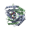
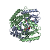
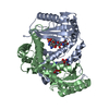
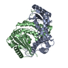
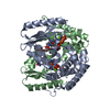
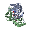
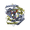
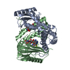
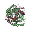
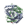
| Title | Wild type ANT bound to neomycin |
|---|
 Components Components | Aminoglycoside NucleotidylTransferase |
|---|
 Keywords Keywords | TRANSFERASE/ANTIBIOTIC / antibiotic resistance / neomycin / TRANSFERASE-ANTIBIOTIC complex |
|---|
| Function / homology |  Function and homology information Function and homology information
nucleotidyltransferase activity / Transferases; Transferring phosphorus-containing groups; Nucleotidyltransferases / response to antibiotic / ATP bindingSimilarity search - Function Kanamycin nucleotidyltransferase, C-terminal / KNTase C-terminal domain / Nucleotidyltransferases domain 2 / Beta Polymerase, domain 2 / Beta Polymerase; domain 2 / Nucleotidyltransferase superfamily / Four Helix Bundle (Hemerythrin (Met), subunit A) / Up-down Bundle / 2-Layer Sandwich / Mainly Alpha / Alpha BetaSimilarity search - Domain/homology |
|---|
| Biological species |   Staphylococcus aureus (bacteria) Staphylococcus aureus (bacteria) |
|---|
| Method |  X-RAY DIFFRACTION / MOLECULAR REPLACEMENT / Resolution: 1.65 Å X-RAY DIFFRACTION / MOLECULAR REPLACEMENT / Resolution: 1.65 Å |
|---|
 Authors Authors | Cuneo, M.J. / Selvaraj, B. |
|---|
 Citation Citation | Journal: ACS Catal / Year: 2020
Title: "Catch and Release": a Variation of the Archetypal NucleotidylTransfer Reaction
Authors: Selvaraj, B. / Kocaman, S. / Trifas, M. / Serpersu, E.H. / Cuneo, M.J. |
|---|
| History | | Deposition | Oct 11, 2019 | Deposition site: RCSB / Processing site: RCSB |
|---|
| Revision 1.0 | May 6, 2020 | Provider: repository / Type: Initial release |
|---|
| Revision 1.1 | Oct 11, 2023 | Group: Data collection / Database references ...Data collection / Database references / Derived calculations / Refinement description
Category: chem_comp_atom / chem_comp_bond ...chem_comp_atom / chem_comp_bond / database_2 / pdbx_initial_refinement_model / pdbx_struct_conn_angle / struct_conn
Item: _database_2.pdbx_DOI / _database_2.pdbx_database_accession ..._database_2.pdbx_DOI / _database_2.pdbx_database_accession / _pdbx_struct_conn_angle.ptnr1_auth_asym_id / _pdbx_struct_conn_angle.ptnr1_auth_comp_id / _pdbx_struct_conn_angle.ptnr1_auth_seq_id / _pdbx_struct_conn_angle.ptnr1_label_asym_id / _pdbx_struct_conn_angle.ptnr1_label_atom_id / _pdbx_struct_conn_angle.ptnr1_label_comp_id / _pdbx_struct_conn_angle.ptnr1_label_seq_id / _pdbx_struct_conn_angle.ptnr3_auth_asym_id / _pdbx_struct_conn_angle.ptnr3_auth_comp_id / _pdbx_struct_conn_angle.ptnr3_auth_seq_id / _pdbx_struct_conn_angle.ptnr3_label_asym_id / _pdbx_struct_conn_angle.ptnr3_label_atom_id / _pdbx_struct_conn_angle.ptnr3_label_comp_id / _pdbx_struct_conn_angle.ptnr3_label_seq_id / _pdbx_struct_conn_angle.value / _struct_conn.pdbx_dist_value / _struct_conn.ptnr1_auth_asym_id / _struct_conn.ptnr1_auth_comp_id / _struct_conn.ptnr1_auth_seq_id / _struct_conn.ptnr1_label_asym_id / _struct_conn.ptnr1_label_atom_id / _struct_conn.ptnr1_label_comp_id / _struct_conn.ptnr1_label_seq_id / _struct_conn.ptnr2_auth_asym_id / _struct_conn.ptnr2_auth_comp_id / _struct_conn.ptnr2_auth_seq_id / _struct_conn.ptnr2_label_asym_id / _struct_conn.ptnr2_label_atom_id / _struct_conn.ptnr2_label_comp_id |
|---|
|
|---|
 Movie
Movie Controller


 Open data
 Basic information
 Structure visualization
 Downloads & links
 Other downloads

 PDBj
PDBj
 Assembly