 Movie
Movie Controller

[English] 日本語
 Yorodumi
- PDB-1jln: Crystal structure of the catalytic domain of protein tyrosine pho... -
+ Open data

- Basic information
| Entry | Database: PDB / ID: 1jln | ||||||
|---|---|---|---|---|---|---|---|
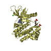
| Title | Crystal structure of the catalytic domain of protein tyrosine phosphatase PTP-SL/BR7 | ||||||
 Components Components | Protein Tyrosine Phosphatase, receptor type, R | ||||||
 Keywords Keywords | HYDROLASE / protein tyrosine phosphatase / PTP-SL / PTPBR7 / ERK2-MAP kinase regulation | ||||||
| Function / homology |  Function and homology information Function and homology informationnegative regulation of epithelial cell migration / ERBB2 signaling pathway / protein-tyrosine-phosphatase / protein tyrosine phosphatase activity / negative regulation of ERK1 and ERK2 cascade / cell junction / in utero embryonic development / protein kinase binding / plasma membrane / cytosol Similarity search - Function | ||||||
| Biological species |  | ||||||
| Method |  X-RAY DIFFRACTION / MOLECULAR REPLACEMENT / Resolution: 1.81 Å X-RAY DIFFRACTION / MOLECULAR REPLACEMENT / Resolution: 1.81 Å | ||||||
 Authors Authors | Szedlacsek, S.E. / Aricescu, A.R. / Fulga, T.A. / Renault, L. / Scheidig, A.J. | ||||||
 Citation Citation | Journal: J.Mol.Biol. / Year: 2001 Title: Crystal structure of PTP-SL/PTPBR7 catalytic domain: implications for MAP kinase regulation. Authors: Szedlacsek, S.E. / Aricescu, A.R. / Fulga, T.A. / Renault, L. / Scheidig, A.J. | ||||||
| History |
|
- Structure visualization
| Structure viewer | Molecule: MolmilJmol/JSmol |
|---|
- Downloads & links
-Download
| PDBx/mmCIF format | 1jln.cif.gz | 76.1 KB | Display | PDBx/mmCIF format |
|---|---|---|---|---|
| PDB format | pdb1jln.ent.gz | 56.1 KB | Display | PDB format |
| PDBx/mmJSON format | 1jln.json.gz | Tree view | PDBx/mmJSON format | |
| Others |  Other downloads Other downloads |
-Validation report
| Summary document | 1jln_validation.pdf.gz | 425.2 KB | Display | wwPDB validaton report |
|---|---|---|---|---|
| Full document | 1jln_full_validation.pdf.gz | 431.8 KB | Display | |
| Data in XML | 1jln_validation.xml.gz | 15.4 KB | Display | |
| Data in CIF | 1jln_validation.cif.gz | 22.2 KB | Display | |
| Arichive directory | https://data.pdbj.org/pub/pdb/validation_reports/jl/1jlnftp://data.pdbj.org/pub/pdb/validation_reports/jl/1jln | HTTPS FTP |
-Related structure data
| Related structure data | 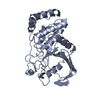 1yfoS S: Starting model for refinement |
|---|---|
| Similar structure data |


-Links
 PDBj
PDBj


- Assembly
| Deposited unit | 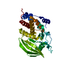
| ||||||||||
|---|---|---|---|---|---|---|---|---|---|---|---|
| 1 |
| ||||||||||
| Unit cell |
|
-Components
| #1: Protein | Mass: 33745.637 Da / Num. of mol.: 1 / Fragment: catalytic domain Source method: isolated from a genetically manipulated source Source: (gene. exp.)  |
|---|---|
| #2: Water | ChemComp-HOH /  Mass: 18.015 Da / Num. of mol.: 238 / Source method: isolated from a natural source / Formula: H2O Mass: 18.015 Da / Num. of mol.: 238 / Source method: isolated from a natural source / Formula: H2O |
-Experimental details
-Experiment
| Experiment | Method: X-RAY DIFFRACTION / Number of used crystals: 1 |
|---|
- Sample preparation
| Crystal | Density Matthews: 2.36 Å3/Da / Density % sol: 47.84 % | |||||||||||||||
|---|---|---|---|---|---|---|---|---|---|---|---|---|---|---|---|---|
| Crystal grow | Temperature: 293 K / Method: vapor diffusion, hanging drop / pH: 7 Details: PEG 3350, pH 7.0, VAPOR DIFFUSION, HANGING DROP at 293K | |||||||||||||||
| Crystal grow | *PLUS Temperature: 20 ℃ | |||||||||||||||
| Components of the solutions | *PLUS
|
-Data collection
| Diffraction | Mean temperature: 100 K |
|---|---|
| Diffraction source | Source: ROTATING ANODE / Type: SIEMENS / Wavelength: 1.5418 Å |
| Detector | Type: SIEMENS HI-STAR / Detector: AREA DETECTOR / Date: Jan 26, 1999 |
| Radiation | Monochromator: graphite / Protocol: SINGLE WAVELENGTH / Monochromatic (M) / Laue (L): M / Scattering type: x-ray |
| Radiation wavelength | Wavelength: 1.5418 Å / Relative weight: 1 |
| Reflection | Resolution: 1.81→19.69 Å / Num. all: 26218 / Num. obs: 24970 / % possible obs: 83.7 % / Observed criterion σ(F): 0 / Observed criterion σ(I): 0 / Redundancy: 4.7 % / Biso Wilson estimate: 28.4 Å2 / Rmerge(I) obs: 0.057 / Rsym value: 5.7 / Net I/σ(I): 17.1 |
| Reflection shell | Resolution: 1.81→1.9 Å / Mean I/σ(I) obs: 2.62 / % possible all: 40.3 |
| Reflection | *PLUS Num. measured all: 118144 |
- Processing
| Software |
| ||||||||||||||||||||||||||||||||||||||||
|---|---|---|---|---|---|---|---|---|---|---|---|---|---|---|---|---|---|---|---|---|---|---|---|---|---|---|---|---|---|---|---|---|---|---|---|---|---|---|---|---|---|
| Refinement | Method to determine structure: MOLECULAR REPLACEMENT Starting model: 1YFO Resolution: 1.81→19.69 Å / Rfactor Rfree error: 0.007 / Data cutoff high absF: 197719.79 / Data cutoff low absF: 0 / Isotropic thermal model: RESTRAINED / Cross valid method: THROUGHOUT / σ(F): 0 / σ(I): 0 / Stereochemistry target values: Engh & Huber
| ||||||||||||||||||||||||||||||||||||||||
| Solvent computation | Solvent model: FLAT MODEL / Bsol: 43.4434 Å2 / ksol: 0.32837 e/Å3 | ||||||||||||||||||||||||||||||||||||||||
| Displacement parameters | Biso mean: 28.3 Å2
| ||||||||||||||||||||||||||||||||||||||||
| Refine analyze |
| ||||||||||||||||||||||||||||||||||||||||
| Refinement step | Cycle: LAST / Resolution: 1.81→19.69 Å
| ||||||||||||||||||||||||||||||||||||||||
| Refine LS restraints |
| ||||||||||||||||||||||||||||||||||||||||
| LS refinement shell | Resolution: 1.8→1.91 Å / Rfactor Rfree error: 0.028 / Total num. of bins used: 6
| ||||||||||||||||||||||||||||||||||||||||
| Xplor file |
| ||||||||||||||||||||||||||||||||||||||||
| Software | *PLUS Name: CNS / Version: 1 / Classification: refinement | ||||||||||||||||||||||||||||||||||||||||
| Refinement | *PLUS σ(F): 0 / % reflection Rfree: 5 % | ||||||||||||||||||||||||||||||||||||||||
| Solvent computation | *PLUS | ||||||||||||||||||||||||||||||||||||||||
| Displacement parameters | *PLUS Biso mean: 28.3 Å2 | ||||||||||||||||||||||||||||||||||||||||
| Refine LS restraints | *PLUS
| ||||||||||||||||||||||||||||||||||||||||
| LS refinement shell | *PLUS Rfactor Rfree: 0.296 / % reflection Rfree: 5 % / Rfactor Rwork: 0.24 |
