 Movie
Movie Controller

[English] 日本語
 Yorodumi
- PDB-1jl0: Structure of a Human S-Adenosylmethionine Decarboxylase Self-proc... -
+ Open data

- Basic information
| Entry | Database: PDB / ID: 1jl0 | ||||||
|---|---|---|---|---|---|---|---|
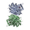
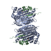
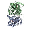
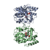
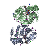
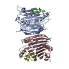
| Title | Structure of a Human S-Adenosylmethionine Decarboxylase Self-processing Ester Intermediate and Mechanism of Putrescine Stimulation of Processing as Revealed by the H243A Mutant | ||||||
 Components Components | S-ADENOSYLMETHIONINE DECARBOXYLASE PROENZYME | ||||||
 Keywords Keywords | LYASE / SPERMIDINE BIOSYNTHESIS / DECARBOXYLASE / PYRUVATE / S-ADENOSYLMETHIONINE / SANDWICH / ALLOSTERIC ENZYME / PYRUVOYL / ESTER INTERMEDIATE / HYDROXYALANINE | ||||||
| Function / homology |  Function and homology information Function and homology informationspermine biosynthetic process / adenosylmethionine decarboxylase / adenosylmethionine decarboxylase activity / Metabolism of polyamines / polyamine metabolic process / putrescine binding / spermidine biosynthetic process / identical protein binding / cytosol Similarity search - Function | ||||||
| Biological species |  Homo sapiens (human) Homo sapiens (human) | ||||||
| Method |  X-RAY DIFFRACTION / SYNCHROTRON / MOLECULAR REPLACEMENT / Resolution: 1.5 Å X-RAY DIFFRACTION / SYNCHROTRON / MOLECULAR REPLACEMENT / Resolution: 1.5 Å | ||||||
 Authors Authors | Ekstrom, J.L. / Tolbert, W.D. / Xiong, H. / Pegg, A.E. / Ealick, S.E. | ||||||
 Citation Citation | Journal: Biochemistry / Year: 2001 Title: Structure of a human S-adenosylmethionine decarboxylase self-processing ester intermediate and mechanism of putrescine stimulation of processing as revealed by the H243A mutant. Authors: Ekstrom, J.L. / Tolbert, W.D. / Xiong, H. / Pegg, A.E. / Ealick, S.E. #1: Journal: Structure / Year: 1999Title: The Crystal Structure of Human S-adenosylmethionine Decarboxylase at 2.25 A Resolution Reveals a Novel Fold Authors: Ekstrom, J.L. / Mathews, I.I. / Stanley, B.A. / Pegg, A.E. / Ealick, S.E. | ||||||
| History |
|
- Structure visualization
| Structure viewer | Molecule: MolmilJmol/JSmol |
|---|
- Downloads & links
-Download
| PDBx/mmCIF format | 1jl0.cif.gz | 166 KB | Display | PDBx/mmCIF format |
|---|---|---|---|---|
| PDB format | pdb1jl0.ent.gz | 130.3 KB | Display | PDB format |
| PDBx/mmJSON format | 1jl0.json.gz | Tree view | PDBx/mmJSON format | |
| Others |  Other downloads Other downloads |
-Validation report
| Arichive directory | https://data.pdbj.org/pub/pdb/validation_reports/jl/1jl0ftp://data.pdbj.org/pub/pdb/validation_reports/jl/1jl0 | HTTPS FTP |
|---|
-Related structure data
| Related structure data |  1jenS S: Starting model for refinement |
|---|---|
| Similar structure data |


-Links
 PDBj
PDBj- Assembly
| Deposited unit | 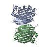
| ||||||||
|---|---|---|---|---|---|---|---|---|---|
| 1 |
| ||||||||
| Unit cell |
| ||||||||
| Details | The enzyme is a dimer and the dimer is in the asymmetric unit. |
-Components
| #1: Protein | Mass: 38298.449 Da / Num. of mol.: 2 / Mutation: H243A Source method: isolated from a genetically manipulated source Source: (gene. exp.) Homo sapiens (human) / Production host:  References: UniProt: P17707, adenosylmethionine decarboxylase #2: Chemical |   Mass: 88.151 Da / Num. of mol.: 2 / Source method: obtained synthetically / Formula: C4H12N2 Mass: 88.151 Da / Num. of mol.: 2 / Source method: obtained synthetically / Formula: C4H12N2#3: Chemical |   Mass: 122.143 Da / Num. of mol.: 2 / Source method: obtained synthetically / Formula: C4H12NO3 / Comment: pH buffer*YM Mass: 122.143 Da / Num. of mol.: 2 / Source method: obtained synthetically / Formula: C4H12NO3 / Comment: pH buffer*YM#4: Water | ChemComp-HOH / |  Mass: 18.015 Da / Num. of mol.: 806 / Source method: isolated from a natural source / Formula: H2O Mass: 18.015 Da / Num. of mol.: 806 / Source method: isolated from a natural source / Formula: H2OHas protein modification | Y | |
|---|
-Experimental details
-Experiment
| Experiment | Method: X-RAY DIFFRACTION / Number of used crystals: 1 |
|---|
- Sample preparation
| Crystal | Density Matthews: 2.32 Å3/Da / Density % sol: 46.97 % | |||||||||||||||
|---|---|---|---|---|---|---|---|---|---|---|---|---|---|---|---|---|
| Crystal grow | Temperature: 277 K / Method: vapor diffusion, hanging drop / pH: 8 Details: PEG 8000, dithiothreitol, tris(hydroxymethyl)aminomethane, pH 8.0, VAPOR DIFFUSION, HANGING DROP, temperature 277K | |||||||||||||||
| Crystal grow | *PLUS Temperature: 4 ℃ | |||||||||||||||
| Components of the solutions | *PLUS
|
-Data collection
| Diffraction source | Source: SYNCHROTRON / Site: APS  / Beamline: 19-ID / Wavelength: 1.03321 / Beamline: 19-ID / Wavelength: 1.03321 |
|---|---|
| Detector | Type: SBC-1 / Detector: CCD / Date: Jun 17, 1999 |
| Radiation | Monochromator: Sagittally focused Si(111) / Protocol: SINGLE WAVELENGTH / Monochromatic (M) / Laue (L): M / Scattering type: x-ray |
| Radiation wavelength | Wavelength: 1.03321 Å / Relative weight: 1 |
| Reflection | Resolution: 1.5→99 Å / Num. all: 111488 / Num. obs: 111488 / % possible obs: 98.8 % / Observed criterion σ(F): 0 / Observed criterion σ(I): 0 / Redundancy: 3.96 % / Biso Wilson estimate: 17.6 Å2 / Rmerge(I) obs: 0.069 / Net I/σ(I): 17.9 |
| Reflection shell | Resolution: 1.5→1.55 Å / Redundancy: 3.92 % / Rmerge(I) obs: 0.275 / Mean I/σ(I) obs: 4.6 / Num. unique all: 11200 / % possible all: 99.5 |
| Reflection | *PLUS |
| Reflection shell | *PLUS Highest resolution: 1.5 Å / % possible obs: 99.5 % |
- Processing
| Software |
| ||||||||||||||||||||||||||||||||||||||||
|---|---|---|---|---|---|---|---|---|---|---|---|---|---|---|---|---|---|---|---|---|---|---|---|---|---|---|---|---|---|---|---|---|---|---|---|---|---|---|---|---|---|
| Refinement | Method to determine structure: MOLECULAR REPLACEMENT Starting model: PDB ENTRY 1JEN Resolution: 1.5→19.91 Å / Rfactor Rfree error: 0.002 / Data cutoff high absF: 472938.91 / Data cutoff low absF: 0 / Isotropic thermal model: RESTRAINED / Cross valid method: THROUGHOUT / σ(F): 0 / Stereochemistry target values: Engh & Huber
| ||||||||||||||||||||||||||||||||||||||||
| Solvent computation | Solvent model: FLAT MODEL / Bsol: 25.7209 Å2 / ksol: 0.306614 e/Å3 | ||||||||||||||||||||||||||||||||||||||||
| Displacement parameters | Biso mean: 16.9 Å2
| ||||||||||||||||||||||||||||||||||||||||
| Refine analyze |
| ||||||||||||||||||||||||||||||||||||||||
| Refinement step | Cycle: LAST / Resolution: 1.5→19.91 Å
| ||||||||||||||||||||||||||||||||||||||||
| Refine LS restraints |
| ||||||||||||||||||||||||||||||||||||||||
| LS refinement shell | Resolution: 1.5→1.59 Å / Rfactor Rfree error: 0.007 / Total num. of bins used: 6
| ||||||||||||||||||||||||||||||||||||||||
| Xplor file |
| ||||||||||||||||||||||||||||||||||||||||
| Software | *PLUS Name: CNS / Version: 1 / Classification: refinement | ||||||||||||||||||||||||||||||||||||||||
| Refinement | *PLUS σ(F): 0 / % reflection Rfree: 10 % / Rfactor obs: 0.215 | ||||||||||||||||||||||||||||||||||||||||
| Solvent computation | *PLUS | ||||||||||||||||||||||||||||||||||||||||
| Displacement parameters | *PLUS Biso mean: 16.9 Å2 | ||||||||||||||||||||||||||||||||||||||||
| Refine LS restraints | *PLUS
| ||||||||||||||||||||||||||||||||||||||||
| LS refinement shell | *PLUS Rfactor Rfree: 0.287 / % reflection Rfree: 9.9 % / Rfactor Rwork: 0.265 |
