Protocol: SINGLE WAVELENGTH / Monochromatic (M) / Laue (L): M
Radiation wavelength
Relative weight: 1
NMR spectrometer
Type: Bruker DMX / Manufacturer: Bruker / Model: DMX / Field strength: 750 MHz
-
Processing
NMR software
Name
Version
Developer
Classification
DYANA
1.5
P. Guentert
refinement
XwinNMR
2.6
Bruker
collection
XEASY
1.3.11
C. Bartels
dataanalysis
Refinement
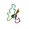
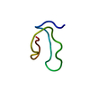
Method: torsion angle dynamics / Software ordinal: 1 Details: Structures are based on a total of 333 distance constraints. This includes 6 upper and 6 lower limits defining 3 disulfide bonds, as well as 3 upper and 3 lower limits defining a peptide ...Details: Structures are based on a total of 333 distance constraints. This includes 6 upper and 6 lower limits defining 3 disulfide bonds, as well as 3 upper and 3 lower limits defining a peptide bond cyclizing the peptide backbone. Residue numbering follows the original description of citation 1, except that for the purposes of structure calculations, the N-terminal residue was taken as Asn8. Therefore, residues 30-36 in this deposition correspond to residues 1-7 in citation 1 and related PDB entry 1kal. Structures were refined in the absence of any artificial constraints defining disulfide bonds until all NOEs had been assigned and low target functions were achieved (tf=0.6). 15 additional calculations were performed with these input data and the inclusion of constraints defining all possible disulfide pairing combinations. The structures containing disulfides between [5(34)-13], [17-29] and [22-27] displayed the lowest target function (0.74, second lowest was 1.54). On the basis of this result and analysis of NOEs observed between Cys sidechain protons, this disulfide bonding arrangement was assumed to be correct and served as the basis for this deposition.
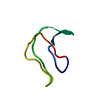
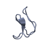
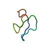
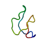
NMR ensemble
Conformer selection criteria: target function / Conformers calculated total number: 50 / Conformers submitted total number: 20
+
About Yorodumi
-
News
-
Feb 9, 2022. New format data for meta-information of EMDB entries
New format data for meta-information of EMDB entries
Version 3 of the EMDB header file is now the official format.
The previous official version 1.9 will be removed from the archive.
In the structure databanks used in Yorodumi, some data are registered as the other names, "COVID-19 virus" and "2019-nCoV". Here are the details of the virus and the list of structure data.
Jan 31, 2019. EMDB accession codes are about to change! (news from PDBe EMDB page)
EMDB accession codes are about to change! (news from PDBe EMDB page)
The allocation of 4 digits for EMDB accession codes will soon come to an end. Whilst these codes will remain in use, new EMDB accession codes will include an additional digit and will expand incrementally as the available range of codes is exhausted. The current 4-digit format prefixed with “EMD-” (i.e. EMD-XXXX) will advance to a 5-digit format (i.e. EMD-XXXXX), and so on. It is currently estimated that the 4-digit codes will be depleted around Spring 2019, at which point the 5-digit format will come into force.
The EM Navigator/Yorodumi systems omit the EMD- prefix.
Related info.:Q: What is EMD? / ID/Accession-code notation in Yorodumi/EM Navigator
Yorodumi is a browser for structure data from EMDB, PDB, SASBDB, etc.
This page is also the successor to EM Navigator detail page, and also detail information page/front-end page for Omokage search.
The word "yorodu" (or yorozu) is an old Japanese word meaning "ten thousand". "mi" (miru) is to see.
Related info.:EMDB / PDB / SASBDB / Comparison of 3 databanks / Yorodumi Search / Aug 31, 2016. New EM Navigator & Yorodumi / Yorodumi Papers / Jmol/JSmol / Function and homology information / Changes in new EM Navigator and Yorodumi
 Movie
Movie Controller


 Open data
 Basic information
 Components
 Keywords
 Function and homology information
 Oldenlandia affinis (plant)
 Authors
 Citation
 Structure visualization
 Downloads & links
 Other downloads


 PDBj
PDBj Assembly
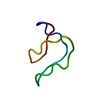
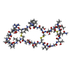
 Type: Cyclic peptide / Class: Antimicrobial, Antitumor / Mass: 2917.345 Da / Num. of mol.: 1 / Source method: isolated from a natural source / Details: extracted from plant / Source: (natural)
  Sample preparation
 Processing