 Movie
Movie Controller


+ Open data

- Basic information
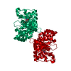
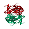
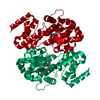
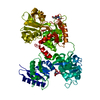
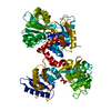
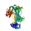
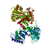
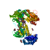
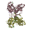
| Entry | Database: PDB / ID: 1jij | ||||||
|---|---|---|---|---|---|---|---|
| Title | Crystal structure of S. aureus TyrRS in complex with SB-239629 | ||||||
 Components Components | tyrosyl-tRNA synthetase | ||||||
 Keywords Keywords | LIGASE / tyrosyl-trna synthetase / staphylococcus aureus / truncation / structure based inhibitor design | ||||||
| Function / homology |  Function and homology information Function and homology informationtyrosyl-tRNA aminoacylation / tyrosine-tRNA ligase / tyrosine-tRNA ligase activity / protein homodimerization activity / RNA binding / ATP binding / cytosol Similarity search - Function | ||||||
| Biological species |   Staphylococcus aureus (bacteria) Staphylococcus aureus (bacteria) | ||||||
| Method |  X-RAY DIFFRACTION / FOURIER SYNTHESIS / Resolution: 3.2 Å X-RAY DIFFRACTION / FOURIER SYNTHESIS / Resolution: 3.2 Å | ||||||
 Authors Authors | Qiu, X. / Janson, C.A. / Smith, W.W. / Jarvest, R.L. | ||||||
 Citation Citation | Journal: Protein Sci. / Year: 2001 Title: Crystal structure of Staphylococcus aureus tyrosyl-tRNA synthetase in complex with a class of potent and specific inhibitors. Authors: Qiu, X. / Janson, C.A. / Smith, W.W. / Green, S.M. / McDevitt, P. / Johanson, K. / Carter, P. / Hibbs, M. / Lewis, C. / Chalker, A. / Fosberry, A. / Lalonde, J. / Berge, J. / Brown, P. / ...Authors: Qiu, X. / Janson, C.A. / Smith, W.W. / Green, S.M. / McDevitt, P. / Johanson, K. / Carter, P. / Hibbs, M. / Lewis, C. / Chalker, A. / Fosberry, A. / Lalonde, J. / Berge, J. / Brown, P. / Houge-Frydrych, C.S. / Jarvest, R.L. | ||||||
| History |
|
- Structure visualization
| Structure viewer | Molecule: MolmilJmol/JSmol |
|---|
- Downloads & links
-Download
| PDBx/mmCIF format | 1jij.cif.gz | 70.4 KB | Display | PDBx/mmCIF format |
|---|---|---|---|---|
| PDB format | pdb1jij.ent.gz | 50.4 KB | Display | PDB format |
| PDBx/mmJSON format | 1jij.json.gz | Tree view | PDBx/mmJSON format | |
| Others |  Other downloads Other downloads |
-Validation report
| Arichive directory | https://data.pdbj.org/pub/pdb/validation_reports/ji/1jijftp://data.pdbj.org/pub/pdb/validation_reports/ji/1jij | HTTPS FTP |
|---|
-Related structure data


-Links
 PDBj
PDBj
- Assembly
| Deposited unit | 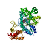
| ||||||||||
|---|---|---|---|---|---|---|---|---|---|---|---|
| 1 | 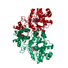
| ||||||||||
| 2 | 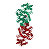
| ||||||||||
| Unit cell |
| ||||||||||
| Details | the biological assembly is a dimer generated from the monomer in ASU and the operation -x,y,-z+1/2 ---- operation 3+1C |
-Components
| #1: Protein | Mass: 47655.449 Da / Num. of mol.: 1 Source method: isolated from a genetically manipulated source Source: (gene. exp.) Staphylococcus aureus (bacteria) / Variant: aureus N315 / Production host: References: GenBank: 13701524, UniProt: A6QHR2*PLUS, tyrosine-tRNA ligase |
|---|---|
| #2: Chemical | ChemComp-629 / [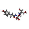  Mass: 415.395 Da / Num. of mol.: 1 / Source method: obtained synthetically / Formula: C17H25N3O9 Mass: 415.395 Da / Num. of mol.: 1 / Source method: obtained synthetically / Formula: C17H25N3O9 |
-Experimental details
-Experiment
| Experiment | Method: X-RAY DIFFRACTION / Number of used crystals: 1 |
|---|
- Sample preparation
| Crystal | Density Matthews: 2.6 Å3/Da / Density % sol: 52.74 % | ||||||||||||||||||||||||||||||
|---|---|---|---|---|---|---|---|---|---|---|---|---|---|---|---|---|---|---|---|---|---|---|---|---|---|---|---|---|---|---|---|
| Crystal grow | Temperature: 298 K / Method: vapor diffusion, sitting drop / pH: 7.25 Details: PEG 1000, CaCl2, pH 7.25, VAPOR DIFFUSION, SITTING DROP at 298K | ||||||||||||||||||||||||||||||
| Crystal grow | *PLUS | ||||||||||||||||||||||||||||||
| Components of the solutions | *PLUS
|
-Data collection
| Diffraction | Mean temperature: 298 K |
|---|---|
| Diffraction source | Source: ROTATING ANODE / Type: RIGAKU RU200 / Wavelength: 1.5418 Å |
| Detector | Type: SIEMENS / Detector: AREA DETECTOR / Date: Jun 6, 1996 |
| Radiation | Protocol: SINGLE WAVELENGTH / Monochromatic (M) / Laue (L): M / Scattering type: x-ray |
| Radiation wavelength | Wavelength: 1.5418 Å / Relative weight: 1 |
| Reflection | Resolution: 3.2→21 Å / Num. all: 8508 / Num. obs: 8399 / % possible obs: 99 % / Observed criterion σ(F): 2 / Observed criterion σ(I): 1 / Redundancy: 3 % / Rmerge(I) obs: 0.108 / Net I/σ(I): 7.1 |
| Reflection shell | Resolution: 3.2→3.22 Å / Redundancy: 3 % / Rmerge(I) obs: 0.288 / Mean I/σ(I) obs: 1.5 / % possible all: 99 |
| Reflection | *PLUS Lowest resolution: 21 Å / % possible obs: 99 % / Num. measured all: 24334 |
| Reflection shell | *PLUS % possible obs: 99 % |
- Processing
| Software |
| ||||||||||||||||||||
|---|---|---|---|---|---|---|---|---|---|---|---|---|---|---|---|---|---|---|---|---|---|
| Refinement | Method to determine structure: FOURIER SYNTHESIS / Resolution: 3.2→8 Å / Cross valid method: THROUGHOUT / σ(F): 2
| ||||||||||||||||||||
| Refinement step | Cycle: LAST / Resolution: 3.2→8 Å
| ||||||||||||||||||||
| Refine LS restraints |
| ||||||||||||||||||||
| Software | *PLUS Name: X-PLOR / Classification: refinement | ||||||||||||||||||||
| Refinement | *PLUS Highest resolution: 3.2 Å / Lowest resolution: 8 Å / σ(F): 2 / % reflection Rfree: 5 % / Rfactor obs: 0.257 | ||||||||||||||||||||
| Solvent computation | *PLUS | ||||||||||||||||||||
| Displacement parameters | *PLUS | ||||||||||||||||||||
| Refine LS restraints | *PLUS Type: x_angle_deg / Dev ideal: 1.9 |
