 Movie
Movie Controller


+ Open data

- Basic information
| Entry | Database: PDB / ID: 1jhn | ||||||
|---|---|---|---|---|---|---|---|
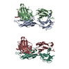
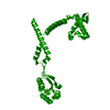
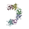
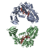
| Title | Crystal Structure of the Lumenal Domain of Calnexin | ||||||
 Components Components | calnexin | ||||||
 Keywords Keywords | CHAPERONE / jelly-roll / beta sandwich | ||||||
| Function / homology |  Function and homology information Function and homology informationmelanosome membrane / clathrin-dependent endocytosis / synaptic vesicle endocytosis / ERAD pathway / mitochondrial membrane / : / carbohydrate binding / protein folding / presynapse / calcium ion binding ...melanosome membrane / clathrin-dependent endocytosis / synaptic vesicle endocytosis / ERAD pathway / mitochondrial membrane / : / carbohydrate binding / protein folding / presynapse / calcium ion binding / endoplasmic reticulum membrane / endoplasmic reticulum Similarity search - Function | ||||||
| Biological species |  | ||||||
| Method |  X-RAY DIFFRACTION / SYNCHROTRON / MIRAS, SAS, MAD / Resolution: 2.9 Å X-RAY DIFFRACTION / SYNCHROTRON / MIRAS, SAS, MAD / Resolution: 2.9 Å | ||||||
 Authors Authors | Schrag, J.D. / Bergeron, J.M. / Li, Y. / Borisova, S. / Hahn, M. / Thomas, D.Y. / Cygler, M. | ||||||
 Citation Citation | Journal: Mol.Cell / Year: 2001 Title: The Structure of calnexin, an ER chaperone involved in quality control of protein folding. Authors: Schrag, J.D. / Bergeron, J.J. / Li, Y. / Borisova, S. / Hahn, M. / Thomas, D.Y. / Cygler, M. #1: Journal: J.Struct.Biol. / Year: 1998Title: Identification and Crystallization of a Protease-Resistant Core of Calnexin that Retains Biological Activity Authors: Hahn, M. / Borisova, S. / Schrag, J.D. / Tessier, D.C. / Zapun, A. / Tom, R. / Kamen, A.A. / Bergeron, J.J. / Thomas, D.Y. / Cygler, M. | ||||||
| History |
|
- Structure visualization
| Structure viewer | Molecule: MolmilJmol/JSmol |
|---|
- Downloads & links
-Download
| PDBx/mmCIF format | 1jhn.cif.gz | 88.4 KB | Display | PDBx/mmCIF format |
|---|---|---|---|---|
| PDB format | pdb1jhn.ent.gz | 64 KB | Display | PDB format |
| PDBx/mmJSON format | 1jhn.json.gz | Tree view | PDBx/mmJSON format | |
| Others |  Other downloads Other downloads |
-Validation report
| Arichive directory | https://data.pdbj.org/pub/pdb/validation_reports/jh/1jhnftp://data.pdbj.org/pub/pdb/validation_reports/jh/1jhn | HTTPS FTP |
|---|
-Related structure data
| Similar structure data |
|---|

-Links
 PDBj
PDBj
- Assembly
| Deposited unit | 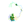
| ||||||||
|---|---|---|---|---|---|---|---|---|---|
| 1 |
| ||||||||
| Unit cell |
|
-Components
| #1: Protein | Mass: 48291.766 Da / Num. of mol.: 1 / Fragment: lumenal domain (residues 45-468) Source method: isolated from a genetically manipulated source Source: (gene. exp.)   Spodoptera frugiperda (fall armyworm) / References: UniProt: P24643 Spodoptera frugiperda (fall armyworm) / References: UniProt: P24643 |
|---|---|
| #2: Chemical | ChemComp-CA /   Mass: 40.078 Da / Num. of mol.: 1 / Source method: obtained synthetically / Formula: Ca Mass: 40.078 Da / Num. of mol.: 1 / Source method: obtained synthetically / Formula: Ca |
| Has protein modification | Y |
-Experimental details
-Experiment
| Experiment | Method: X-RAY DIFFRACTION / Number of used crystals: 1 |
|---|
- Sample preparation
| Crystal | Density Matthews: 2.73 Å3/Da / Density % sol: 54.94 % | ||||||||||||||||||||||||||||||||||||||||||||||||||||||||||||
|---|---|---|---|---|---|---|---|---|---|---|---|---|---|---|---|---|---|---|---|---|---|---|---|---|---|---|---|---|---|---|---|---|---|---|---|---|---|---|---|---|---|---|---|---|---|---|---|---|---|---|---|---|---|---|---|---|---|---|---|---|---|
| Crystal grow | Temperature: 277 K / Method: vapor diffusion, hanging drop / pH: 7.5 Details: HEPES, MPD, Calcium chloride, pH 7.5, VAPOR DIFFUSION, HANGING DROP, temperature 277K | ||||||||||||||||||||||||||||||||||||||||||||||||||||||||||||
| Crystal grow | *PLUS Temperature: 4 ℃Details: used microseeding, Hahn, M., (1998) J. Struct. Biol., 123, 260. | ||||||||||||||||||||||||||||||||||||||||||||||||||||||||||||
| Components of the solutions | *PLUS
|
-Data collection
| Diffraction | Mean temperature: 100 K |
|---|---|
| Diffraction source | Source: SYNCHROTRON / Site: NSLS  / Beamline: X8C / Wavelength: 1.4 Å / Beamline: X8C / Wavelength: 1.4 Å |
| Detector | Type: ADSC QUANTUM 4 / Detector: CCD / Date: Jan 27, 1999 |
| Radiation | Monochromator: Si 111 / Protocol: SINGLE WAVELENGTH / Monochromatic (M) / Laue (L): M / Scattering type: x-ray |
| Radiation wavelength | Wavelength: 1.4 Å / Relative weight: 1 |
| Reflection | Resolution: 2.9→35 Å / Num. all: 12339 / Num. obs: 12339 / % possible obs: 99.4 % / Observed criterion σ(F): 0 / Observed criterion σ(I): 0 / Redundancy: 6.7 % / Biso Wilson estimate: 84.5 Å2 / Rmerge(I) obs: 0.046 / Rsym value: 0.046 / Net I/σ(I): 23.4 |
| Reflection shell | Resolution: 2.9→3 Å / Redundancy: 6.7 % / Rmerge(I) obs: 0.245 / Mean I/σ(I) obs: 5.4 / Rsym value: 0.245 / % possible all: 99.6 |
| Reflection | *PLUS Num. measured all: 82347 |
| Reflection shell | *PLUS % possible obs: 99.6 % |
- Processing
| Software |
| ||||||||||||||||||||
|---|---|---|---|---|---|---|---|---|---|---|---|---|---|---|---|---|---|---|---|---|---|
| Refinement | Method to determine structure: MIRAS, SAS, MAD / Resolution: 2.9→33.8 Å / σ(F): 4 / σ(I): 2 / Stereochemistry target values: Engh and Huber / Details: phase restraints used
| ||||||||||||||||||||
| Displacement parameters | Biso mean: 59.5 Å2
| ||||||||||||||||||||
| Refine analyze |
| ||||||||||||||||||||
| Refinement step | Cycle: LAST / Resolution: 2.9→33.8 Å
| ||||||||||||||||||||
| Refine LS restraints |
| ||||||||||||||||||||
| Software | *PLUS Name: CNS / Classification: refinement | ||||||||||||||||||||
| Refinement | *PLUS Highest resolution: 2.9 Å / Lowest resolution: 33.8 Å / σ(F): 4 / Rfactor obs: 0.327 | ||||||||||||||||||||
| Solvent computation | *PLUS | ||||||||||||||||||||
| Displacement parameters | *PLUS Biso mean: 59.5 Å2 | ||||||||||||||||||||
| Refine LS restraints | *PLUS
|
