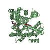
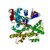
 Movie
Movie Controller


+ Open data

- Basic information
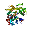
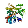
| Entry | Database: PDB / ID: 1jcf | ||||||
|---|---|---|---|---|---|---|---|
| Title | MREB FROM THERMOTOGA MARITIMA, TRIGONAL | ||||||
 Components Components | ROD SHAPE-DETERMINING PROTEIN MREB | ||||||
 Keywords Keywords | STRUCTURAL PROTEIN / MreB / rod-shape determining / Mbl / actin / hsp-70 / FtsZ | ||||||
| Function / homology |  Function and homology information Function and homology information | ||||||
| Biological species |   Thermotoga maritima (bacteria) Thermotoga maritima (bacteria) | ||||||
| Method |  X-RAY DIFFRACTION / SYNCHROTRON / MOLECULAR REPLACEMENT / Resolution: 2.1 Å X-RAY DIFFRACTION / SYNCHROTRON / MOLECULAR REPLACEMENT / Resolution: 2.1 Å | ||||||
 Authors Authors | van den Ent, F. / Amos, L.A. / Lowe, J. | ||||||
 Citation Citation | Journal: Nature / Year: 2001 Title: Prokaryotic origin of the actin cytoskeleton. Authors: van den Ent, F. / Amos, L.A. / Lowe, J. | ||||||
| History |
|
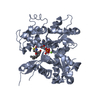
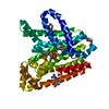
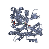
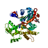
- Structure visualization
| Structure viewer | Molecule: MolmilJmol/JSmol |
|---|
- Downloads & links
-Download
| PDBx/mmCIF format | 1jcf.cif.gz | 82 KB | Display | PDBx/mmCIF format |
|---|---|---|---|---|
| PDB format | pdb1jcf.ent.gz | 60.9 KB | Display | PDB format |
| PDBx/mmJSON format | 1jcf.json.gz | Tree view | PDBx/mmJSON format | |
| Others |  Other downloads Other downloads |
-Validation report
| Summary document | 1jcf_validation.pdf.gz | 425 KB | Display | wwPDB validaton report |
|---|---|---|---|---|
| Full document | 1jcf_full_validation.pdf.gz | 429.2 KB | Display | |
| Data in XML | 1jcf_validation.xml.gz | 17.2 KB | Display | |
| Data in CIF | 1jcf_validation.cif.gz | 25.6 KB | Display | |
| Arichive directory | https://data.pdbj.org/pub/pdb/validation_reports/jc/1jcfftp://data.pdbj.org/pub/pdb/validation_reports/jc/1jcf | HTTPS FTP |
-Related structure data
| Related structure data | 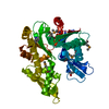 1jceSC 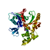 1jcgC S: Starting model for refinement C: citing same article ( |
|---|---|
| Similar structure data |


-Links
 PDBj
PDBj
- Assembly
| Deposited unit | 
| ||||||||
|---|---|---|---|---|---|---|---|---|---|
| 1 |
| ||||||||
| Unit cell |
| ||||||||
| Details | Crystal packing generates one-dimensional filaments alon a and b, similar to one strand in F-actin (protofilament) |
-Components
| #1: Protein | Mass: 36799.582 Da / Num. of mol.: 1 Source method: isolated from a genetically manipulated source Source: (gene. exp.) Thermotoga maritima (bacteria) / Gene: TM0588 / Plasmid: PHIS17 / Production host: |
|---|---|
| #2: Water | ChemComp-HOH /  Mass: 18.015 Da / Num. of mol.: 318 / Source method: isolated from a natural source / Formula: H2O Mass: 18.015 Da / Num. of mol.: 318 / Source method: isolated from a natural source / Formula: H2O |
-Experimental details
-Experiment
| Experiment | Method: X-RAY DIFFRACTION / Number of used crystals: 1 |
|---|
- Sample preparation
| Crystal | Density Matthews: 3 Å3/Da / Density % sol: 58.98 % | ||||||||||||||||||||||||||||||
|---|---|---|---|---|---|---|---|---|---|---|---|---|---|---|---|---|---|---|---|---|---|---|---|---|---|---|---|---|---|---|---|
| Crystal grow | *PLUS Temperature: 19 ℃ / pH: 10.5 / Method: vapor diffusion, sitting drop | ||||||||||||||||||||||||||||||
| Components of the solutions | *PLUS
|
-Data collection
| Diffraction | Mean temperature: 100 K |
|---|---|
| Diffraction source | Source: SYNCHROTRON / Site: ESRF  / Beamline: ID14-4 / Wavelength: 0.9393 Å / Beamline: ID14-4 / Wavelength: 0.9393 Å |
| Detector | Type: ADSC QUANTUM 4 / Detector: CCD / Date: May 8, 2001 |
| Radiation | Monochromator: double / Protocol: SINGLE WAVELENGTH / Monochromatic (M) / Laue (L): M / Scattering type: x-ray |
| Radiation wavelength | Wavelength: 0.9393 Å / Relative weight: 1 |
| Reflection | Resolution: 2.1→50 Å / Num. all: 27249 / Num. obs: 26139 / % possible obs: 95.8 % / Observed criterion σ(F): 0 / Observed criterion σ(I): 0 / Redundancy: 3.1 % / Biso Wilson estimate: 25 Å2 / Rmerge(I) obs: 0.06 / Net I/σ(I): 13.7 |
| Reflection shell | Resolution: 2.09→2.2 Å / Redundancy: 2.7 % / Rmerge(I) obs: 0.115 / % possible all: 92.4 |
| Reflection | *PLUS Rmerge(I) obs: 0.06 |
- Processing
| Software |
| |||||||||||||||||||||||||
|---|---|---|---|---|---|---|---|---|---|---|---|---|---|---|---|---|---|---|---|---|---|---|---|---|---|---|
| Refinement | Method to determine structure: MOLECULAR REPLACEMENT Starting model: PDB ENTRY 1JCE Resolution: 2.1→50 Å / σ(F): 0 / σ(I): 0 / Stereochemistry target values: CNS 1.0, protein_rep.param
| |||||||||||||||||||||||||
| Displacement parameters | Biso mean: 30.31 Å2 | |||||||||||||||||||||||||
| Refinement step | Cycle: LAST / Resolution: 2.1→50 Å
| |||||||||||||||||||||||||
| Refine LS restraints |
| |||||||||||||||||||||||||
| Software | *PLUS Name: CNS / Version: 1 / Classification: refinement | |||||||||||||||||||||||||
| Refinement | *PLUS Highest resolution: 2.1 Å / Lowest resolution: 50 Å / σ(F): 0 | |||||||||||||||||||||||||
| Solvent computation | *PLUS | |||||||||||||||||||||||||
| Displacement parameters | *PLUS |
