 Movie
Movie Controller

[English] 日本語
 Yorodumi
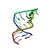
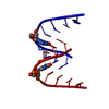
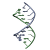
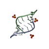
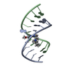
- PDB-1jb8: The Crystal Structure of an RNA/DNA Hybrid Reveals Novel Intermol... -
+ Open data

- Basic information
| Entry | Database: PDB / ID: 1jb8 | ||||||
|---|---|---|---|---|---|---|---|
| Title | The Crystal Structure of an RNA/DNA Hybrid Reveals Novel Intermolecular Intercalation | ||||||
 Components Components |
| ||||||
 Keywords Keywords | DNA/RNA / DNA/DNA intercalation / RNA/DNA hybrid / RNA/DNA polypurine tract / DNA-RNA COMPLEX | ||||||
| Function / homology | DNA / RNA Function and homology information Function and homology information | ||||||
| Method |  X-RAY DIFFRACTION / MOLECULAR REPLACEMENT / Resolution: 2.38 Å X-RAY DIFFRACTION / MOLECULAR REPLACEMENT / Resolution: 2.38 Å | ||||||
 Authors Authors | Han, G.W. / Kopka, M.L. / Langs, D. / Dickerson, R.E. | ||||||
 Citation Citation | Journal: Proc.Natl.Acad.Sci.USA / Year: 2003 Title: Crystal structure of an RNADNA hybrid reveals intermolecular intercalation: Dimer formation by base-pair swapping Authors: Han, G.W. / Kopka, M.L. / Langs, D. / Sawaya, M.R. / Dickerson, R.E. | ||||||
| History |
|
- Structure visualization
| Structure viewer | Molecule: MolmilJmol/JSmol |
|---|
- Downloads & links
-Download
| PDBx/mmCIF format | 1jb8.cif.gz | 21.5 KB | Display | PDBx/mmCIF format |
|---|---|---|---|---|
| PDB format | pdb1jb8.ent.gz | 13.4 KB | Display | PDB format |
| PDBx/mmJSON format | 1jb8.json.gz | Tree view | PDBx/mmJSON format | |
| Others |  Other downloads Other downloads |
-Validation report
| Arichive directory | https://data.pdbj.org/pub/pdb/validation_reports/jb/1jb8ftp://data.pdbj.org/pub/pdb/validation_reports/jb/1jb8 | HTTPS FTP |
|---|
-Related structure data
| Similar structure data | |
|---|---|
| Other databases |
|


-Links
 PDBj
PDBj- Assembly
| Deposited unit | 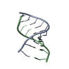
| ||||||||
|---|---|---|---|---|---|---|---|---|---|
| 1 |
| ||||||||
| Unit cell |
| ||||||||
| Components on special symmetry positions |
|
-Components
| #1: RNA chain | Mass: 3255.076 Da / Num. of mol.: 1 / Source method: obtained synthetically / Details: RNA/DNA hybrid of PPT sequence of HIV |
|---|---|
| #2: DNA chain | Mass: 2991.961 Da / Num. of mol.: 1 / Source method: obtained synthetically Details: Synthesized by the solid-phase phosphoramidate method on an Eppendorf ECOSYN D300 Synthesizer. |
| #3: Water | ChemComp-HOH /  Mass: 18.015 Da / Num. of mol.: 66 / Source method: isolated from a natural source / Formula: H2O Mass: 18.015 Da / Num. of mol.: 66 / Source method: isolated from a natural source / Formula: H2O |
-Experimental details
-Experiment
| Experiment | Method: X-RAY DIFFRACTION / Number of used crystals: 1 |
|---|
- Sample preparation
| Crystal | Density Matthews: 2.38 Å3/Da / Density % sol: 48.24 % | |||||||||||||||||||||||||
|---|---|---|---|---|---|---|---|---|---|---|---|---|---|---|---|---|---|---|---|---|---|---|---|---|---|---|
| Crystal grow | Temperature: 277 K / Method: vapor diffusion, hanging drop Details: MPD, magnesium acetate, spermidine, Na cacodylate, VAPOR DIFFUSION, HANGING DROP, temperature 277K | |||||||||||||||||||||||||
| Components of the solutions |
| |||||||||||||||||||||||||
| Crystal grow | *PLUS pH: 5.8 / Method: vapor diffusion, sitting drop | |||||||||||||||||||||||||
| Components of the solutions | *PLUS
|
-Data collection
| Diffraction | Mean temperature: 93 K |
|---|---|
| Diffraction source | Source: ROTATING ANODE / Type: ENRAF-NONIUS / Wavelength: 1.5418 Å |
| Detector | Type: RIGAKU RAXIS IV / Detector: IMAGE PLATE / Date: Aug 15, 2000 / Details: mirrors |
| Radiation | Protocol: SINGLE WAVELENGTH / Monochromatic (M) / Laue (L): M / Scattering type: x-ray |
| Radiation wavelength | Wavelength: 1.5418 Å / Relative weight: 1 |
| Reflection | Resolution: 2.38→10 Å / Num. all: 18386 / Num. obs: 2814 / % possible obs: 98.9 % / Observed criterion σ(I): 2 / Redundancy: 6.5 % / Rmerge(I) obs: 0.053 |
| Reflection shell | Resolution: 2.3→2.38 Å / Rmerge(I) obs: 0.78 / % possible all: 96.9 |
- Processing
| Software |
| ||||||||||||||||||||||||||||||
|---|---|---|---|---|---|---|---|---|---|---|---|---|---|---|---|---|---|---|---|---|---|---|---|---|---|---|---|---|---|---|---|
| Refinement | Method to determine structure: MOLECULAR REPLACEMENT Starting model: P2(1)2(1)2(1) structure of the same sequence of RNA/DNA hybrid Resolution: 2.38→10 Å / Isotropic thermal model: Isotropic / σ(F): 2
| ||||||||||||||||||||||||||||||
| Refinement step | Cycle: LAST / Resolution: 2.38→10 Å
| ||||||||||||||||||||||||||||||
| Refine LS restraints |
| ||||||||||||||||||||||||||||||
| LS refinement shell |
|
