 Movie
Movie Controller


+ Open data

- Basic information
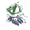
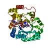
| Entry | Database: PDB / ID: 1jb5 | ||||||
|---|---|---|---|---|---|---|---|
| Title | CRYSTAL STRUCTURE OF NTF2 M118E MUTANT | ||||||
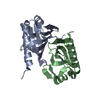
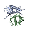
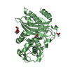
 Components Components | NUCLEAR TRANSPORT FACTOR 2 | ||||||
 Keywords Keywords | PROTEIN TRANSPORT / NTF2 / Transport | ||||||
| Function / homology |  Function and homology information Function and homology informationprotein localization to nuclear pore / negative regulation of vascular endothelial growth factor production / nuclear pore central transport channel / structural constituent of nuclear pore / nuclear inner membrane / nuclear import signal receptor activity / nuclear outer membrane / mRNA transport / protein export from nucleus / positive regulation of protein import into nucleus ...protein localization to nuclear pore / negative regulation of vascular endothelial growth factor production / nuclear pore central transport channel / structural constituent of nuclear pore / nuclear inner membrane / nuclear import signal receptor activity / nuclear outer membrane / mRNA transport / protein export from nucleus / positive regulation of protein import into nucleus / protein import into nucleus / small GTPase binding / nuclear membrane / nucleoplasm / identical protein binding / cytosol Similarity search - Function | ||||||
| Biological species |  | ||||||
| Method |  X-RAY DIFFRACTION / SYNCHROTRON / MOLECULAR REPLACEMENT / Resolution: 2.3 Å X-RAY DIFFRACTION / SYNCHROTRON / MOLECULAR REPLACEMENT / Resolution: 2.3 Å | ||||||
 Authors Authors | Chaillan-Huntington, C. / Butler, P.J. / Huntington, J.A. / Akin, D. / Feldherr, C. / Stewart, M. | ||||||
 Citation Citation | Journal: J.Mol.Biol. / Year: 2001 Title: NTF2 monomer-dimer equilibrium. Authors: Chaillan-Huntington, C. / Butler, P.J. / Huntington, J.A. / Akin, D. / Feldherr, C. / Stewart, M. | ||||||
| History |
|
- Structure visualization
| Structure viewer | Molecule: MolmilJmol/JSmol |
|---|
- Downloads & links
-Download
| PDBx/mmCIF format | 1jb5.cif.gz | 61.8 KB | Display | PDBx/mmCIF format |
|---|---|---|---|---|
| PDB format | pdb1jb5.ent.gz | 46.1 KB | Display | PDB format |
| PDBx/mmJSON format | 1jb5.json.gz | Tree view | PDBx/mmJSON format | |
| Others |  Other downloads Other downloads |
-Validation report
| Arichive directory | https://data.pdbj.org/pub/pdb/validation_reports/jb/1jb5ftp://data.pdbj.org/pub/pdb/validation_reports/jb/1jb5 | HTTPS FTP |
|---|
-Related structure data
| Related structure data |  1jb2C 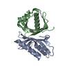 1jb4SC C: citing same article ( S: Starting model for refinement |
|---|---|
| Similar structure data |


-Links
 PDBj
PDBj
- Assembly
| Deposited unit | 
| ||||||||
|---|---|---|---|---|---|---|---|---|---|
| 1 |
| ||||||||
| Unit cell |
|
-Components
| #1: Protein | Mass: 14475.319 Da / Num. of mol.: 2 / Mutation: M118E Source method: isolated from a genetically manipulated source Source: (gene. exp.)  #2: Water | ChemComp-HOH / |  Mass: 18.015 Da / Num. of mol.: 112 / Source method: isolated from a natural source / Formula: H2O Mass: 18.015 Da / Num. of mol.: 112 / Source method: isolated from a natural source / Formula: H2O |
|---|
-Experimental details
-Experiment
| Experiment | Method: X-RAY DIFFRACTION / Number of used crystals: 1 |
|---|
- Sample preparation
| Crystal | Density Matthews: 1.88 Å3/Da / Density % sol: 34.5 % | |||||||||||||||||||||||||||||||||||
|---|---|---|---|---|---|---|---|---|---|---|---|---|---|---|---|---|---|---|---|---|---|---|---|---|---|---|---|---|---|---|---|---|---|---|---|---|
| Crystal grow | Temperature: 291 K / Method: vapor diffusion, hanging drop / pH: 4.5 Details: PEG, pH 4.5, VAPOR DIFFUSION, HANGING DROP, temperature 291K | |||||||||||||||||||||||||||||||||||
| Crystal grow | *PLUS | |||||||||||||||||||||||||||||||||||
| Components of the solutions | *PLUS
|
-Data collection
| Diffraction | Mean temperature: 100 K |
|---|---|
| Diffraction source | Source: SYNCHROTRON / Site: SRS  / Beamline: PX7.2 / Wavelength: 1.488 Å / Beamline: PX7.2 / Wavelength: 1.488 Å |
| Detector | Type: MARRESEARCH / Detector: IMAGE PLATE |
| Radiation | Protocol: SINGLE WAVELENGTH / Monochromatic (M) / Laue (L): M / Scattering type: x-ray |
| Radiation wavelength | Wavelength: 1.488 Å / Relative weight: 1 |
| Reflection | Resolution: 2.3→30 Å / Num. all: 9197 / Num. obs: 9197 / % possible obs: 96.2 % / Observed criterion σ(F): 0 / Observed criterion σ(I): 0 / Redundancy: 2.5 % / Rmerge(I) obs: 0.098 / Net I/σ(I): 9.6 |
| Reflection shell | Resolution: 2.3→2.38 Å / Redundancy: 2 % / Rmerge(I) obs: 0.272 / % possible all: 96.2 |
| Reflection | *PLUS Highest resolution: 2.3 Å / Lowest resolution: 30 Å / Num. measured all: 73003 |
| Reflection shell | *PLUS Highest resolution: 2.3 Å / % possible obs: 96.2 % / Mean I/σ(I) obs: 2.4 |
- Processing
| Software |
| |||||||||||||||||||||||||
|---|---|---|---|---|---|---|---|---|---|---|---|---|---|---|---|---|---|---|---|---|---|---|---|---|---|---|
| Refinement | Method to determine structure: MOLECULAR REPLACEMENT Starting model: 1JB4 Resolution: 2.3→30 Å / σ(F): 0 / σ(I): 0 / Stereochemistry target values: Ergh & Huber
| |||||||||||||||||||||||||
| Displacement parameters | Biso mean: 28 Å2 | |||||||||||||||||||||||||
| Refinement step | Cycle: LAST / Resolution: 2.3→30 Å
| |||||||||||||||||||||||||
| Refine LS restraints |
| |||||||||||||||||||||||||
| Refinement | *PLUS Highest resolution: 2.3 Å / Lowest resolution: 30 Å / σ(F): 0 / % reflection Rfree: 10 % | |||||||||||||||||||||||||
| Solvent computation | *PLUS | |||||||||||||||||||||||||
| Displacement parameters | *PLUS Biso mean: 28 Å2 | |||||||||||||||||||||||||
| Refine LS restraints | *PLUS Type: x_angle_deg / Dev ideal: 1.3 |
