 Movie
Movie Controller


+ Open data

- Basic information
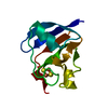
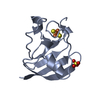
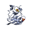
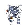
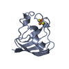
| Entry | Database: PDB / ID: 1j7c | |||||||||
|---|---|---|---|---|---|---|---|---|---|---|
| Title | STRUCTURE OF THE ANABAENA FERREDOXIN MUTANT E95K | |||||||||
 Components Components | FERREDOXIN I | |||||||||
 Keywords Keywords | ELECTRON TRANSPORT / iron-sulfur / ferredoxin | |||||||||
| Function / homology |  Function and homology information Function and homology informationelectron transport chain / 2 iron, 2 sulfur cluster binding / electron transfer activity / metal ion binding Similarity search - Function | |||||||||
| Biological species |  Nostoc sp. PCC (bacteria) Nostoc sp. PCC (bacteria) | |||||||||
| Method |  X-RAY DIFFRACTION / FOURIER SYNTHESIS / Resolution: 1.8 Å X-RAY DIFFRACTION / FOURIER SYNTHESIS / Resolution: 1.8 Å | |||||||||
 Authors Authors | Hurley, J.K. / Weber-Main, A.M. / Stankovich, M.T. / Benning, M.M. / Thoden, J.B. / Vanhooke, J.L. / Holden, H.M. / Chae, Y.K. / Xia, B. / Cheng, H. ...Hurley, J.K. / Weber-Main, A.M. / Stankovich, M.T. / Benning, M.M. / Thoden, J.B. / Vanhooke, J.L. / Holden, H.M. / Chae, Y.K. / Xia, B. / Cheng, H. / Markley, J.L. / Martinez-Julvez, M. / Gomez-Moreno, C. / Schmeits, J.L. / Tollin, G. | |||||||||
 Citation Citation | Journal: Biochemistry / Year: 1997 Title: Structure-function relationships in Anabaena ferredoxin: correlations between X-ray crystal structures, reduction potentials, and rate constants of electron transfer to ferredoxin:NADP+ ...Title: Structure-function relationships in Anabaena ferredoxin: correlations between X-ray crystal structures, reduction potentials, and rate constants of electron transfer to ferredoxin:NADP+ reductase for site-specific ferredoxin mutants. Authors: Hurley, J.K. / Weber-Main, A.M. / Stankovich, M.T. / Benning, M.M. / Thoden, J.B. / Vanhooke, J.L. / Holden, H.M. / Chae, Y.K. / Xia, B. / Cheng, H. / Markley, J.L. / Martinez-Julvez, M. / ...Authors: Hurley, J.K. / Weber-Main, A.M. / Stankovich, M.T. / Benning, M.M. / Thoden, J.B. / Vanhooke, J.L. / Holden, H.M. / Chae, Y.K. / Xia, B. / Cheng, H. / Markley, J.L. / Martinez-Julvez, M. / Gomez-Moreno, C. / Schmeits, J.L. / Tollin, G. | |||||||||
| History |
|
- Structure visualization
| Structure viewer | Molecule: MolmilJmol/JSmol |
|---|
- Downloads & links
-Download
| PDBx/mmCIF format | 1j7c.cif.gz | 33.6 KB | Display | PDBx/mmCIF format |
|---|---|---|---|---|
| PDB format | pdb1j7c.ent.gz | 22.2 KB | Display | PDB format |
| PDBx/mmJSON format | 1j7c.json.gz | Tree view | PDBx/mmJSON format | |
| Others |  Other downloads Other downloads |
-Validation report
| Arichive directory | https://data.pdbj.org/pub/pdb/validation_reports/j7/1j7cftp://data.pdbj.org/pub/pdb/validation_reports/j7/1j7c | HTTPS FTP |
|---|
-Related structure data


-Links
 PDBj
PDBj


- Assembly
| Deposited unit | 
| ||||||||
|---|---|---|---|---|---|---|---|---|---|
| 1 |
| ||||||||
| Unit cell |
|
-Components
| #1: Protein | Mass: 10705.690 Da / Num. of mol.: 1 / Mutation: E95K Source method: isolated from a genetically manipulated source Source: (gene. exp.) Nostoc sp. PCC (bacteria) / Strain: 7120 / Plasmid: PAN662 / Production host: |
|---|---|
| #2: Chemical | ChemComp-FES /   Mass: 175.820 Da / Num. of mol.: 1 / Source method: obtained synthetically / Formula: Fe2S2 Mass: 175.820 Da / Num. of mol.: 1 / Source method: obtained synthetically / Formula: Fe2S2 |
| #3: Water | ChemComp-HOH /  Mass: 18.015 Da / Num. of mol.: 93 / Source method: isolated from a natural source / Formula: H2O Mass: 18.015 Da / Num. of mol.: 93 / Source method: isolated from a natural source / Formula: H2O |
-Experimental details
-Experiment
| Experiment | Method: X-RAY DIFFRACTION / Number of used crystals: 1 |
|---|
- Sample preparation
| Crystal | Density Matthews: 2.69 Å3/Da / Density % sol: 54.32 % | ||||||||||||||||||||
|---|---|---|---|---|---|---|---|---|---|---|---|---|---|---|---|---|---|---|---|---|---|
| Crystal grow | Temperature: 277 K / Method: vapor diffusion, hanging drop / pH: 5.5 Details: ammonium sulfate, potassium succinate, 2-methyl-2,4-pentanediol, pH 5.5, VAPOR DIFFUSION, HANGING DROP, temperature 277K | ||||||||||||||||||||
| Crystal grow | *PLUS Temperature: 4 ℃ | ||||||||||||||||||||
| Components of the solutions | *PLUS
|
-Data collection
| Diffraction | Mean temperature: 277 K |
|---|---|
| Diffraction source | Source: ROTATING ANODE / Type: RIGAKU RU200 / Wavelength: 1.5418 Å |
| Detector | Type: SIEMENS HI-STAR / Detector: AREA DETECTOR / Date: Dec 12, 1996 / Details: supper mirrors |
| Radiation | Monochromator: supper mirrors / Protocol: SINGLE WAVELENGTH / Monochromatic (M) / Laue (L): M / Scattering type: x-ray |
| Radiation wavelength | Wavelength: 1.5418 Å / Relative weight: 1 |
| Reflection | Resolution: 1.8→30 Å / Num. all: 10573 / Num. obs: 10573 / % possible obs: 89 % / Observed criterion σ(F): 0 / Observed criterion σ(I): 0 / Redundancy: 2.7 % / Rmerge(I) obs: 0.036 / Net I/σ(I): 21.5 |
| Reflection shell | Resolution: 1.8→1.86 Å / Redundancy: 1.8 % / Rmerge(I) obs: 0.14 / Mean I/σ(I) obs: 3.4 / % possible all: 74 |
| Reflection | *PLUS % possible obs: 89 % / Num. measured all: 24013 |
- Processing
| Software |
| ||||||||||||
|---|---|---|---|---|---|---|---|---|---|---|---|---|---|
| Refinement | Method to determine structure: FOURIER SYNTHESIS / Resolution: 1.8→30 Å / σ(F): 0 / σ(I): 0 / Stereochemistry target values: Engh & Huber /
| ||||||||||||
| Refinement step | Cycle: LAST / Resolution: 1.8→30 Å
| ||||||||||||
| Refine LS restraints |
| ||||||||||||
| Software | *PLUS Name: TNT / Classification: refinement | ||||||||||||
| Refinement | *PLUS σ(F): 0 / Rfactor all: 0.174 | ||||||||||||
| Solvent computation | *PLUS | ||||||||||||
| Displacement parameters | *PLUS | ||||||||||||
| Refine LS restraints | *PLUS Type: t_angle_deg / Dev ideal: 2.12 |
