 Movie
Movie Controller

[English] 日本語
 Yorodumi
- PDB-1j6u: Crystal structure of UDP-N-acetylmuramate-alanine ligase MurC (TM... -
+ Open data

- Basic information
| Entry | Database: PDB / ID: 1j6u | ||||||
|---|---|---|---|---|---|---|---|
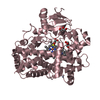
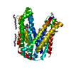
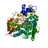
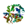
| Title | Crystal structure of UDP-N-acetylmuramate-alanine ligase MurC (TM0231) from Thermotoga maritima at 2.3 A resolution | ||||||
 Components Components | UDP-N-acetylmuramate-alanine ligase MurC | ||||||
 Keywords Keywords | LIGASE / STRUCTURAL GENOMICS / TM0231 / UDP-N-acetylmuramate-alanine ligase MurC / JCSG / PSI / Protein Structure Initiative / Joint Center for Structural Genomics | ||||||
| Function / homology |  Function and homology information Function and homology informationUDP-N-acetylmuramate-L-alanine ligase / UDP-N-acetylmuramate-L-alanine ligase activity / peptidoglycan biosynthetic process / cell wall organization / regulation of cell shape / cell division / ATP binding / cytoplasm Similarity search - Function | ||||||
| Biological species |   Thermotoga maritima (bacteria) Thermotoga maritima (bacteria) | ||||||
| Method |  X-RAY DIFFRACTION / SYNCHROTRON / MOLECULAR REPLACEMENT / Resolution: 2.3 Å X-RAY DIFFRACTION / SYNCHROTRON / MOLECULAR REPLACEMENT / Resolution: 2.3 Å | ||||||
 Authors Authors | Joint Center for Structural Genomics (JCSG) | ||||||
 Citation Citation | Journal: Proteins / Year: 2004 Title: Crystal structure of an Udp-n-acetylmuramate-alanine ligase MurC (TM0231) from Thermotoga maritima at 2.3 A resolution. Authors: Spraggon, G. / Schwarzenbacher, R. / Kreusch, A. / Lee, C.C. / Abdubek, P. / Ambing, E. / Biorac, T. / Brinen, L.S. / Canaves, J.M. / Cambell, J. / Chiu, H.J. / Dai, X. / Deacon, A.M. / ...Authors: Spraggon, G. / Schwarzenbacher, R. / Kreusch, A. / Lee, C.C. / Abdubek, P. / Ambing, E. / Biorac, T. / Brinen, L.S. / Canaves, J.M. / Cambell, J. / Chiu, H.J. / Dai, X. / Deacon, A.M. / DiDonato, M. / Elsliger, M.A. / Eshagi, S. / Floyd, R. / Godzik, A. / Grittini, C. / Grzechnik, S.K. / Hampton, E. / Jaroszewski, L. / Karlak, C. / Klock, H.E. / Koesema, E. / Kovarik, J.S. / Kuhn, P. / Levin, I. / McMullan, D. / McPhillips, T.M. / Miller, M.D. / Morse, A. / Moy, K. / Ouyang, J. / Page, R. / Quijano, K. / Robb, A. / Stevens, R.C. / van den Bedem, H. / Velasquez, J. / Vincent, J. / von Delft, F. / Wang, X. / West, B. / Wolf, G. / Xu, Q. / Hodgson, K.O. / Wooley, J. / Lesley, S.A. / Wilson, I.A. | ||||||
| History |
|
- Structure visualization
| Structure viewer | Molecule: MolmilJmol/JSmol |
|---|
- Downloads & links
-Download
| PDBx/mmCIF format | 1j6u.cif.gz | 107.3 KB | Display | PDBx/mmCIF format |
|---|---|---|---|---|
| PDB format | pdb1j6u.ent.gz | 81.6 KB | Display | PDB format |
| PDBx/mmJSON format | 1j6u.json.gz | Tree view | PDBx/mmJSON format | |
| Others |  Other downloads Other downloads |
-Validation report
| Arichive directory | https://data.pdbj.org/pub/pdb/validation_reports/j6/1j6uftp://data.pdbj.org/pub/pdb/validation_reports/j6/1j6u | HTTPS FTP |
|---|
-Related structure data
| Similar structure data | |
|---|---|
| Other databases |


-Links
 PDBj
PDBj- Assembly
| Deposited unit | 
| ||||||||
|---|---|---|---|---|---|---|---|---|---|
| 1 |
| ||||||||
| Unit cell |
| ||||||||
| Components on special symmetry positions |
|
-Components
| #1: Protein | Mass: 53827.219 Da / Num. of mol.: 1 Source method: isolated from a genetically manipulated source Source: (gene. exp.) Thermotoga maritima (bacteria) / Gene: TM0231 / Production host: References: UniProt: Q9WY73, UDP-N-acetylmuramate-L-alanine ligase |
|---|---|
| #2: Water | ChemComp-HOH /  Mass: 18.015 Da / Num. of mol.: 245 / Source method: isolated from a natural source / Formula: H2O Mass: 18.015 Da / Num. of mol.: 245 / Source method: isolated from a natural source / Formula: H2O |
| Has protein modification | Y |
-Experimental details
-Experiment
| Experiment | Method: X-RAY DIFFRACTION / Number of used crystals: 1 |
|---|
- Sample preparation
| Crystal | Density Matthews: 5.58 Å3/Da / Density % sol: 78.82 % |
|---|---|
| Crystal grow | Temperature: 293 K / pH: 7.5 Details: 1.4M sodium potassium phosphate ph 7.5, VAPOR DIFFUSION,SITTING DROP,NANODROP, temperature 293K, pH 7.50 |
| Crystal grow | *PLUS Method: otherDetails: Santarsiero, B.D., (2002) J. Appl. Crystallogr., 35, 278. |
-Data collection
| Diffraction | Mean temperature: 100 K |
|---|---|
| Diffraction source | Source: SYNCHROTRON / Site: ALS  / Beamline: 5.0.3 / Wavelength: 1 / Beamline: 5.0.3 / Wavelength: 1 |
| Detector | Type: ADSC QUANTUM 4 / Detector: CCD / Date: Feb 1, 2002 |
| Radiation | Monochromator: ASYMMETRICALLY CUT SI CRYSTAL, CYLINDRICALLY BENT Protocol: SINGLE WAVELENGTH / Monochromatic (M) / Laue (L): M / Scattering type: x-ray |
| Radiation wavelength | Wavelength: 1 Å / Relative weight: 1 |
| Reflection | Resolution: 2.3→182 Å / Num. obs: 52273 / % possible obs: 95.3 % / Redundancy: 4.3 % / Biso Wilson estimate: 55.2 Å2 / Rsym value: 0.099 / Net I/σ(I): 9.55 |
| Reflection shell | Resolution: 2.28→2.37 Å / Redundancy: 4.6 % / Mean I/σ(I) obs: 0.96 / Rsym value: 0.77 / % possible all: 97.8 |
| Reflection | *PLUS Highest resolution: 2.3 Å / Lowest resolution: 70.71 Å / Num. obs: 54912 / % possible obs: 97.7 % / Redundancy: 4.3 % / Num. measured all: 252363 / Rmerge(I) obs: 0.124 |
| Reflection shell | *PLUS Highest resolution: 2.3 Å / % possible obs: 96.5 % / Rmerge(I) obs: 0.55 / Mean I/σ(I) obs: 1.6 |
- Processing
| Software |
| ||||||||||||||||||||||||||||||||||||||||||||||||||||||||||||||||||||||||||||||||
|---|---|---|---|---|---|---|---|---|---|---|---|---|---|---|---|---|---|---|---|---|---|---|---|---|---|---|---|---|---|---|---|---|---|---|---|---|---|---|---|---|---|---|---|---|---|---|---|---|---|---|---|---|---|---|---|---|---|---|---|---|---|---|---|---|---|---|---|---|---|---|---|---|---|---|---|---|---|---|---|---|---|
| Refinement | Method to determine structure: MOLECULAR REPLACEMENT / Resolution: 2.3→39.6 Å / Isotropic thermal model: ISOTROPIC / Cross valid method: FREE R / σ(F): 0 Stereochemistry target values: STANDARD CNS DICTIONARY/ENGH AND HUBER
| ||||||||||||||||||||||||||||||||||||||||||||||||||||||||||||||||||||||||||||||||
| Solvent computation | Solvent model: BULK SOLVENT CORRECTION / Bsol: 0.4 Å2 / ksol: 89.02 e/Å3 | ||||||||||||||||||||||||||||||||||||||||||||||||||||||||||||||||||||||||||||||||
| Displacement parameters | Biso mean: 49.26 Å2
| ||||||||||||||||||||||||||||||||||||||||||||||||||||||||||||||||||||||||||||||||
| Refine analyze |
| ||||||||||||||||||||||||||||||||||||||||||||||||||||||||||||||||||||||||||||||||
| Refinement step | Cycle: LAST / Resolution: 2.3→39.6 Å
| ||||||||||||||||||||||||||||||||||||||||||||||||||||||||||||||||||||||||||||||||
| Refine LS restraints |
| ||||||||||||||||||||||||||||||||||||||||||||||||||||||||||||||||||||||||||||||||
| LS refinement shell | Resolution: 2.3→2.32 Å / Total num. of bins used: 50
| ||||||||||||||||||||||||||||||||||||||||||||||||||||||||||||||||||||||||||||||||
| Refinement | *PLUS Num. reflection obs: 54912 | ||||||||||||||||||||||||||||||||||||||||||||||||||||||||||||||||||||||||||||||||
| Solvent computation | *PLUS | ||||||||||||||||||||||||||||||||||||||||||||||||||||||||||||||||||||||||||||||||
| Displacement parameters | *PLUS |
