 Movie
Movie Controller

[English] 日本語
 Yorodumi
- PDB-1j4a: INSIGHTS INTO DOMAIN CLOSURE, SUBSTRATE SPECIFICITY AND CATALYSIS... -
+ Open data

- Basic information
| Entry | Database: PDB / ID: 1j4a | ||||||
|---|---|---|---|---|---|---|---|
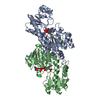
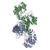
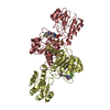
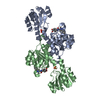
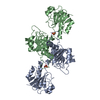
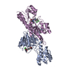
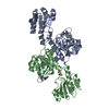
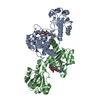
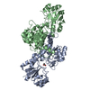
| Title | INSIGHTS INTO DOMAIN CLOSURE, SUBSTRATE SPECIFICITY AND CATALYSIS OF D-LACTATE DEHYDROGENASE FROM LACTOBACILLUS BULGARICUS | ||||||
 Components Components | D-LACTATE DEHYDROGENASE | ||||||
 Keywords Keywords | OXIDOREDUCTASE / NAD-DEPENDENT DEHYDROGENASE / REVERSIBLE INTERCONVERSION OF PYRUVATE INTO D-LACTATE | ||||||
| Function / homology |  Function and homology information Function and homology informationD-lactate dehydrogenase / D-lactate dehydrogenase (NAD+) activity / lactate metabolic process / NAD binding Similarity search - Function | ||||||
| Biological species |  Lactobacillus delbrueckii subsp. bulgaricus (bacteria) Lactobacillus delbrueckii subsp. bulgaricus (bacteria) | ||||||
| Method |  X-RAY DIFFRACTION / SYNCHROTRON / MOLECULAR REPLACEMENT / Resolution: 1.9 Å X-RAY DIFFRACTION / SYNCHROTRON / MOLECULAR REPLACEMENT / Resolution: 1.9 Å | ||||||
 Authors Authors | Razeto, A. / Kochhar, S. / Hottinger, H. / Dauter, M. / Wilson, K.S. / Lamzin, V.S. | ||||||
 Citation Citation | Journal: J.Mol.Biol. / Year: 2002 Title: Domain closure, substrate specificity and catalysis of D-lactate dehydrogenase from Lactobacillus bulgaricus. Authors: Razeto, A. / Kochhar, S. / Hottinger, H. / Dauter, M. / Wilson, K.S. / Lamzin, V.S. #1: Journal: Thesis / Year: 1999Title: Lysine Replacing Histidine in the Acid-Base Catalysis of D-Lactate Dehydrogenase: Structural Investigation on Enzymatic Stereoselectivity. Authors: Razeto, A. | ||||||
| History |
|
- Structure visualization
| Structure viewer | Molecule: MolmilJmol/JSmol |
|---|
- Downloads & links
-Download
| PDBx/mmCIF format | 1j4a.cif.gz | 291.7 KB | Display | PDBx/mmCIF format |
|---|---|---|---|---|
| PDB format | pdb1j4a.ent.gz | 234.6 KB | Display | PDB format |
| PDBx/mmJSON format | 1j4a.json.gz | Tree view | PDBx/mmJSON format | |
| Others |  Other downloads Other downloads |
-Validation report
| Arichive directory | https://data.pdbj.org/pub/pdb/validation_reports/j4/1j4aftp://data.pdbj.org/pub/pdb/validation_reports/j4/1j4a | HTTPS FTP |
|---|
-Related structure data


-Links
 PDBj
PDBj- Assembly
| Deposited unit | 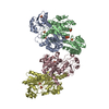
| ||||||||||||
|---|---|---|---|---|---|---|---|---|---|---|---|---|---|
| 1 | 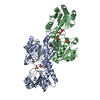
| ||||||||||||
| 2 | 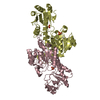
| ||||||||||||
| Unit cell |
| ||||||||||||
| Noncrystallographic symmetry (NCS) | NCS oper:
|
-Components
| #1: Protein | Mass: 37112.160 Da / Num. of mol.: 4 / Mutation: H297K Source method: isolated from a genetically manipulated source Source: (gene. exp.) Lactobacillus delbrueckii subsp. bulgaricus (bacteria)Species: Lactobacillus delbrueckii / Strain: N42 / Cellular location: CYTOPLASM / Gene: LDHD / Plasmid: PMD19 / Cellular location (production host): CYTOPLASM / Gene (production host): LDHD / Production host: #2: Chemical | ChemComp-SO4 /   Mass: 96.063 Da / Num. of mol.: 11 / Source method: obtained synthetically / Formula: SO4 Mass: 96.063 Da / Num. of mol.: 11 / Source method: obtained synthetically / Formula: SO4#3: Water | ChemComp-HOH / |  Mass: 18.015 Da / Num. of mol.: 900 / Source method: isolated from a natural source / Formula: H2O Mass: 18.015 Da / Num. of mol.: 900 / Source method: isolated from a natural source / Formula: H2O |
|---|
-Experimental details
-Experiment
| Experiment | Method: X-RAY DIFFRACTION / Number of used crystals: 1 |
|---|
- Sample preparation
| Crystal | Density Matthews: 2.91 Å3/Da / Density % sol: 57.4 % | ||||||||||||||||||||||||
|---|---|---|---|---|---|---|---|---|---|---|---|---|---|---|---|---|---|---|---|---|---|---|---|---|---|
| Crystal grow | pH: 6.5 Details: Orthorhombic crystals were grown by vapour diffusion from solutions of 20 % PEG 6K, 0.2 M ammonium sulphate in 0.1 M cacodylate buffer pH 6.5. | ||||||||||||||||||||||||
| Crystal grow | *PLUS Method: vapor diffusion | ||||||||||||||||||||||||
| Components of the solutions | *PLUS
|
-Data collection
| Diffraction | Mean temperature: 100 K |
|---|---|
| Diffraction source | Source: SYNCHROTRON / Site: EMBL/DESY, HAMBURG  / Beamline: X11 / Wavelength: 0.906 / Beamline: X11 / Wavelength: 0.906 |
| Detector | Type: MARRESEARCH / Detector: IMAGE PLATE / Date: Dec 1, 1998 / Details: MIRRORS |
| Radiation | Monochromator: SILICON CRYSTAL / Protocol: SINGLE WAVELENGTH / Monochromatic (M) / Laue (L): M / Scattering type: x-ray |
| Radiation wavelength | Wavelength: 0.906 Å / Relative weight: 1 |
| Reflection | Resolution: 1.9→19 Å / Num. obs: 132348 / % possible obs: 98.4 % / Observed criterion σ(I): -3 / Redundancy: 3.1 % / Biso Wilson estimate: 0.215 Å2 / Rmerge(I) obs: 0.047 / Net I/σ(I): 16.1 |
| Reflection shell | Resolution: 1.9→1.93 Å / Rmerge(I) obs: 0.327 / Mean I/σ(I) obs: 2.2 / % possible all: 92.4 |
| Reflection | *PLUS Rmerge(I) obs: 0.047 |
| Reflection shell | *PLUS % possible obs: 92.4 % / Rmerge(I) obs: 0.327 |
- Processing
| Software |
| ||||||||||||||||||||||||||||||||||||||||||||||||||||||||||||||||||||||||||||||||||||
|---|---|---|---|---|---|---|---|---|---|---|---|---|---|---|---|---|---|---|---|---|---|---|---|---|---|---|---|---|---|---|---|---|---|---|---|---|---|---|---|---|---|---|---|---|---|---|---|---|---|---|---|---|---|---|---|---|---|---|---|---|---|---|---|---|---|---|---|---|---|---|---|---|---|---|---|---|---|---|---|---|---|---|---|---|---|
| Refinement | Method to determine structure: MOLECULAR REPLACEMENT Starting model: MODEL OF NATIVE D-LDH WHICH IS CURRENTLY BEING DEPOSITED IN THE PDB Resolution: 1.9→19 Å / SU B: 2.44 / Cross valid method: THROUGHOUT APART FROM THE LAST 3 CYCLES / σ(F): 0 / ESU R Free: 0.15 Details: THE DENSITY OF THE CATALYTIC DOMAIN OF SUBUNIT C IS VERY POOR: 49 RESIDUES WERE ASSIGNED ZERO OCCUPANCY. IN ALL SUBUNITS THERE IS NO DENSITY FOR THE METHIONINE AT THE N-TERMINUS AND IN ...Details: THE DENSITY OF THE CATALYTIC DOMAIN OF SUBUNIT C IS VERY POOR: 49 RESIDUES WERE ASSIGNED ZERO OCCUPANCY. IN ALL SUBUNITS THERE IS NO DENSITY FOR THE METHIONINE AT THE N-TERMINUS AND IN SUBUNITS A,B,C FOR GLY-333 AT THE C-TERMINUS.
| ||||||||||||||||||||||||||||||||||||||||||||||||||||||||||||||||||||||||||||||||||||
| Displacement parameters | Biso mean: 0.327 Å2 | ||||||||||||||||||||||||||||||||||||||||||||||||||||||||||||||||||||||||||||||||||||
| Refinement step | Cycle: LAST / Resolution: 1.9→19 Å
| ||||||||||||||||||||||||||||||||||||||||||||||||||||||||||||||||||||||||||||||||||||
| Refine LS restraints |
| ||||||||||||||||||||||||||||||||||||||||||||||||||||||||||||||||||||||||||||||||||||
| Refinement | *PLUS % reflection Rfree: 5 % / Rfactor obs: 0.204 / Rfactor Rfree: 0.259 / Rfactor Rwork: 0.204 | ||||||||||||||||||||||||||||||||||||||||||||||||||||||||||||||||||||||||||||||||||||
| Solvent computation | *PLUS | ||||||||||||||||||||||||||||||||||||||||||||||||||||||||||||||||||||||||||||||||||||
| Displacement parameters | *PLUS |
