 Movie
Movie Controller

[English] 日本語
 Yorodumi
- PDB-1j49: INSIGHTS INTO DOMAIN CLOSURE, SUBSTRATE SPECIFICITY AND CATALYSIS... -
+ Open data

- Basic information
| Entry | Database: PDB / ID: 1j49 | ||||||
|---|---|---|---|---|---|---|---|
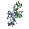
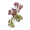
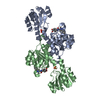
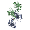
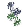
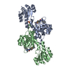
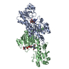
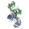
| Title | INSIGHTS INTO DOMAIN CLOSURE, SUBSTRATE SPECIFICITY AND CATALYSIS OF D-LACTATE DEHYDROGENASE FROM LACTOBACILLUS BULGARICUS | ||||||
 Components Components | D-LACTATE DEHYDROGENASE | ||||||
 Keywords Keywords | OXIDOREDUCTASE / NAD-DEPENDENT DEHYDROGENASE / LAST STEP OF GLYCOLYSIS UNDER ANAEROBIC CONDITIONS / REVERSIBLE INTERCONVERSION OF PYRUVATE INTO D-LACTATE | ||||||
| Function / homology |  Function and homology information Function and homology informationD-lactate dehydrogenase / D-lactate dehydrogenase (NAD+) activity / lactate metabolic process / NAD binding Similarity search - Function | ||||||
| Biological species |  Lactobacillus delbrueckii subsp. bulgaricus (bacteria) Lactobacillus delbrueckii subsp. bulgaricus (bacteria) | ||||||
| Method |  X-RAY DIFFRACTION / SYNCHROTRON / MOLECULAR REPLACEMENT / Resolution: 2.2 Å X-RAY DIFFRACTION / SYNCHROTRON / MOLECULAR REPLACEMENT / Resolution: 2.2 Å | ||||||
 Authors Authors | Razeto, A. / Kochhar, S. / Hottinger, H. / Dauter, M. / Wilson, K.S. / Lamzin, V.S. | ||||||
 Citation Citation | Journal: J.Mol.Biol. / Year: 2002 Title: Domain closure, substrate specificity and catalysis of D-lactate dehydrogenase from Lactobacillus bulgaricus. Authors: Razeto, A. / Kochhar, S. / Hottinger, H. / Dauter, M. / Wilson, K.S. / Lamzin, V.S. | ||||||
| History |
| ||||||
| Remark 999 | SEQUENCE The authors maintain that their sequence is correct. The density for these residues ...SEQUENCE The authors maintain that their sequence is correct. The density for these residues suggests ILE59, ARG117, AND VAL152 instead of those in the database reference match. |
- Structure visualization
| Structure viewer | Molecule: MolmilJmol/JSmol |
|---|
- Downloads & links
-Download
| PDBx/mmCIF format | 1j49.cif.gz | 149.3 KB | Display | PDBx/mmCIF format |
|---|---|---|---|---|
| PDB format | pdb1j49.ent.gz | 118.2 KB | Display | PDB format |
| PDBx/mmJSON format | 1j49.json.gz | Tree view | PDBx/mmJSON format | |
| Others |  Other downloads Other downloads |
-Validation report
| Arichive directory | https://data.pdbj.org/pub/pdb/validation_reports/j4/1j49ftp://data.pdbj.org/pub/pdb/validation_reports/j4/1j49 | HTTPS FTP |
|---|
-Related structure data
| Related structure data |  1j4aC  2dldS S: Starting model for refinement C: citing same article ( |
|---|---|
| Similar structure data |

-Links
 PDBj
PDBj- Assembly
| Deposited unit | 
| ||||||||
|---|---|---|---|---|---|---|---|---|---|
| 1 |
| ||||||||
| Unit cell |
| ||||||||
| Noncrystallographic symmetry (NCS) | NCS oper: (Code: given Matrix: (-0.31073, -0.06203, 0.94847), Vector: |
-Components
| #1: Protein | Mass: 37121.125 Da / Num. of mol.: 2 Source method: isolated from a genetically manipulated source Source: (gene. exp.) Lactobacillus delbrueckii subsp. bulgaricus (bacteria)Species: Lactobacillus delbrueckii / Strain: N42 Description: NESTLE CULTURE COLLECTION. PCR AMPLIFIED LDHA GENE Cellular location: CYTOPLASM / Gene: LDHA / Plasmid: PKBULDH / Cellular location (production host): CYTOPLASM / Gene (production host): LDHA / Production host: #2: Chemical |   Mass: 663.425 Da / Num. of mol.: 2 / Source method: obtained synthetically / Formula: C21H27N7O14P2 / Comment: NAD*YM Mass: 663.425 Da / Num. of mol.: 2 / Source method: obtained synthetically / Formula: C21H27N7O14P2 / Comment: NAD*YM#3: Chemical | ChemComp-SO4 / |   Mass: 96.063 Da / Num. of mol.: 1 / Source method: obtained synthetically / Formula: SO4 Mass: 96.063 Da / Num. of mol.: 1 / Source method: obtained synthetically / Formula: SO4#4: Water | ChemComp-HOH / |  Mass: 18.015 Da / Num. of mol.: 279 / Source method: isolated from a natural source / Formula: H2O Mass: 18.015 Da / Num. of mol.: 279 / Source method: isolated from a natural source / Formula: H2O |
|---|
-Experimental details
-Experiment
| Experiment | Method: X-RAY DIFFRACTION / Number of used crystals: 2 |
|---|
- Sample preparation
| Crystal | Density Matthews: 2.45 Å3/Da / Density % sol: 49 % | ||||||||||||||||||||||||||||||
|---|---|---|---|---|---|---|---|---|---|---|---|---|---|---|---|---|---|---|---|---|---|---|---|---|---|---|---|---|---|---|---|
| Crystal grow | pH: 5 / Details: pH 5.0 | ||||||||||||||||||||||||||||||
| Crystal grow | *PLUS Method: vapor diffusion, hanging drop | ||||||||||||||||||||||||||||||
| Components of the solutions | *PLUS
|
-Data collection
| Diffraction | Mean temperature: 298 K |
|---|---|
| Diffraction source | Source: SYNCHROTRON / Site: EMBL/DESY, HAMBURG  / Beamline: X11 / Wavelength: 0.916 / Beamline: X11 / Wavelength: 0.916 |
| Detector | Type: MARRESEARCH / Detector: IMAGE PLATE / Date: Dec 1, 1994 / Details: MIRRORS |
| Radiation | Monochromator: SILICON CRYSTAL / Protocol: SINGLE WAVELENGTH / Monochromatic (M) / Laue (L): M / Scattering type: x-ray |
| Radiation wavelength | Wavelength: 0.916 Å / Relative weight: 1 |
| Reflection | Resolution: 2.2→15 Å / Num. obs: 38147 / % possible obs: 98.7 % / Observed criterion σ(I): 2 / Redundancy: 5.8 % / Biso Wilson estimate: 36.1 Å2 / Rmerge(I) obs: 0.071 / Net I/σ(I): 19.4 |
| Reflection shell | Resolution: 2.2→2.24 Å / Redundancy: 6.82 % / Rmerge(I) obs: 0.21 / Mean I/σ(I) obs: 3.73 / % possible all: 93.3 |
| Reflection | *PLUS Lowest resolution: 15 Å / Rmerge(I) obs: 0.071 |
| Reflection shell | *PLUS % possible obs: 93.3 % / Rmerge(I) obs: 0.21 / Mean I/σ(I) obs: 3.7 |
- Processing
| Software |
| ||||||||||||||||||||||||||||||||||||||||||||||||||||||||||||||||||||||||||||||||||||
|---|---|---|---|---|---|---|---|---|---|---|---|---|---|---|---|---|---|---|---|---|---|---|---|---|---|---|---|---|---|---|---|---|---|---|---|---|---|---|---|---|---|---|---|---|---|---|---|---|---|---|---|---|---|---|---|---|---|---|---|---|---|---|---|---|---|---|---|---|---|---|---|---|---|---|---|---|---|---|---|---|---|---|---|---|---|
| Refinement | Method to determine structure: MOLECULAR REPLACEMENT Starting model: PDB ENTRY 2DLD Resolution: 2.2→15 Å / SU B: 4.35 / SU ML: 0.11 Cross valid method: THROUGHOUT (EXCEPT FOR THE LAST 2 REFINEMENT CYCLES) σ(F): 0 / ESU R: 0.27 / ESU R Free: 0.24
| ||||||||||||||||||||||||||||||||||||||||||||||||||||||||||||||||||||||||||||||||||||
| Displacement parameters | Biso mean: 44.4 Å2 | ||||||||||||||||||||||||||||||||||||||||||||||||||||||||||||||||||||||||||||||||||||
| Refinement step | Cycle: LAST / Resolution: 2.2→15 Å
| ||||||||||||||||||||||||||||||||||||||||||||||||||||||||||||||||||||||||||||||||||||
| Refine LS restraints |
| ||||||||||||||||||||||||||||||||||||||||||||||||||||||||||||||||||||||||||||||||||||
| Refinement | *PLUS % reflection Rfree: 5 % / Rfactor obs: 0.209 / Rfactor Rfree: 0.271 / Rfactor Rwork: 0.209 | ||||||||||||||||||||||||||||||||||||||||||||||||||||||||||||||||||||||||||||||||||||
| Solvent computation | *PLUS | ||||||||||||||||||||||||||||||||||||||||||||||||||||||||||||||||||||||||||||||||||||
| Displacement parameters | *PLUS |
