 Movie
Movie Controller

[English] 日本語
 Yorodumi
- PDB-1j3b: Crystal structure of ATP-dependent phosphoenolpyruvate carboxykin... -
+ Open data

- Basic information
| Entry | Database: PDB / ID: 1j3b | ||||||
|---|---|---|---|---|---|---|---|
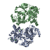
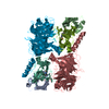
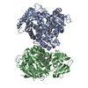
| Title | Crystal structure of ATP-dependent phosphoenolpyruvate carboxykinase from Thermus thermophilus HB8 | ||||||
 Components Components | ATP-dependent phosphoenolpyruvate carboxykinase | ||||||
 Keywords Keywords | TRANSFERASE / phosphoenolpyruvate carboxykinase / adenosine triphosphate / Thermus thermophilus / RIKEN Structural Genomics/Proteomics Initiative / RSGI / Structural Genomics | ||||||
| Function / homology |  Function and homology information Function and homology informationphosphoenolpyruvate carboxykinase (ATP) / phosphoenolpyruvate carboxykinase (ATP) activity / gluconeogenesis / ATP binding / metal ion binding / cytosol Similarity search - Function | ||||||
| Biological species |   Thermus thermophilus (bacteria) Thermus thermophilus (bacteria) | ||||||
| Method |  X-RAY DIFFRACTION / SYNCHROTRON / MOLECULAR REPLACEMENT / Resolution: 2 Å X-RAY DIFFRACTION / SYNCHROTRON / MOLECULAR REPLACEMENT / Resolution: 2 Å | ||||||
 Authors Authors | Sugahara, M. / Miyano, M. / Kunishima, N. / RIKEN Structural Genomics/Proteomics Initiative (RSGI) | ||||||
 Citation Citation | Journal: Acta Crystallogr.,Sect.D / Year: 2005 Title: Structure of ATP-dependent phosphoenolpyruvate carboxykinase from Thermus thermophilus HB8 showing the structural basis of induced fit and thermostability. Authors: Sugahara, M. / Ohshima, N. / Ukita, Y. / Sugahara, M. / Kunishima, N. | ||||||
| History |
|
- Structure visualization
| Structure viewer | Molecule: MolmilJmol/JSmol |
|---|
- Downloads & links
-Download
| PDBx/mmCIF format | 1j3b.cif.gz | 229.7 KB | Display | PDBx/mmCIF format |
|---|---|---|---|---|
| PDB format | pdb1j3b.ent.gz | 182.9 KB | Display | PDB format |
| PDBx/mmJSON format | 1j3b.json.gz | Tree view | PDBx/mmJSON format | |
| Others |  Other downloads Other downloads |
-Validation report
| Arichive directory | https://data.pdbj.org/pub/pdb/validation_reports/j3/1j3bftp://data.pdbj.org/pub/pdb/validation_reports/j3/1j3b | HTTPS FTP |
|---|
-Related structure data
| Related structure data | 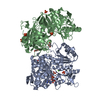 1xkvC 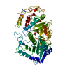 1oenS S: Starting model for refinement C: citing same article ( |
|---|---|
| Similar structure data | |
| Other databases |


-Links
 PDBj
PDBj- Assembly
| Deposited unit | 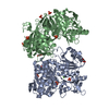
| ||||||||
|---|---|---|---|---|---|---|---|---|---|
| 1 |
| ||||||||
| Unit cell |
| ||||||||
| Details | The biological assembly is a dimer in the asymmetric unit. |
-Components
| #1: Protein | Mass: 59387.191 Da / Num. of mol.: 2 Source method: isolated from a genetically manipulated source Source: (gene. exp.) Thermus thermophilus (bacteria) / Strain: HB8 / Plasmid: pET11a / Production host: #2: Chemical |   Mass: 40.078 Da / Num. of mol.: 2 / Source method: obtained synthetically / Formula: Ca Mass: 40.078 Da / Num. of mol.: 2 / Source method: obtained synthetically / Formula: Ca#3: Chemical | ChemComp-PO4 /   Mass: 94.971 Da / Num. of mol.: 9 / Source method: obtained synthetically / Formula: PO4 Mass: 94.971 Da / Num. of mol.: 9 / Source method: obtained synthetically / Formula: PO4#4: Chemical |   Mass: 92.094 Da / Num. of mol.: 2 / Source method: obtained synthetically / Formula: C3H8O3 Mass: 92.094 Da / Num. of mol.: 2 / Source method: obtained synthetically / Formula: C3H8O3#5: Water | ChemComp-HOH / |  Mass: 18.015 Da / Num. of mol.: 709 / Source method: isolated from a natural source / Formula: H2O Mass: 18.015 Da / Num. of mol.: 709 / Source method: isolated from a natural source / Formula: H2O |
|---|
-Experimental details
-Experiment
| Experiment | Method: X-RAY DIFFRACTION / Number of used crystals: 1 |
|---|
- Sample preparation
| Crystal | Density Matthews: 2.9 Å3/Da / Density % sol: 57.25 % | ||||||||||||||||||||||||
|---|---|---|---|---|---|---|---|---|---|---|---|---|---|---|---|---|---|---|---|---|---|---|---|---|---|
| Crystal grow | Temperature: 291 K / Method: microbatch / pH: 6.3 Details: potassium phosphate, sodium phosphate, pH 6.3, MICROBATCH, temperature 291K | ||||||||||||||||||||||||
| Crystal grow | *PLUS Method: batch method | ||||||||||||||||||||||||
| Components of the solutions | *PLUS
|
-Data collection
| Diffraction | Mean temperature: 100 K |
|---|---|
| Diffraction source | Source: SYNCHROTRON / Site: SPring-8  / Beamline: BL38B1 / Wavelength: 1 Å / Beamline: BL38B1 / Wavelength: 1 Å |
| Detector | Type: ADSC QUANTUM 4 / Detector: CCD / Date: Oct 2, 2002 / Details: mirrors |
| Radiation | Monochromator: mirrors / Protocol: SINGLE WAVELENGTH / Monochromatic (M) / Laue (L): M / Scattering type: x-ray |
| Radiation wavelength | Wavelength: 1 Å / Relative weight: 1 |
| Reflection | Resolution: 2→50 Å / Num. all: 99163 / Num. obs: 99163 / % possible obs: 99.9 % / Observed criterion σ(F): 0 / Observed criterion σ(I): 0 / Redundancy: 7.32 % / Biso Wilson estimate: 33.1 Å2 / Rmerge(I) obs: 0.074 / Rsym value: 0.07 / Net I/σ(I): 8 |
| Reflection shell | Resolution: 2→2.07 Å / Redundancy: 7.13 % / Rmerge(I) obs: 0.713 / Mean I/σ(I) obs: 4.19 / Num. unique all: 9804 / Rsym value: 0.675 / % possible all: 100 |
| Reflection | *PLUS Lowest resolution: 50 Å / Num. measured all: 726185 |
| Reflection shell | *PLUS Highest resolution: 2 Å / % possible obs: 100 % / Mean I/σ(I) obs: 4.2 |
- Processing
| Software |
| |||||||||||||||||||||||||
|---|---|---|---|---|---|---|---|---|---|---|---|---|---|---|---|---|---|---|---|---|---|---|---|---|---|---|
| Refinement | Method to determine structure: MOLECULAR REPLACEMENT Starting model: PDB entry 1OEN Resolution: 2→36.1 Å / Isotropic thermal model: Anisotropic / Cross valid method: THROUGHOUT / σ(F): 0 / Stereochemistry target values: Engh & Huber
| |||||||||||||||||||||||||
| Displacement parameters | Biso mean: 36.6 Å2
| |||||||||||||||||||||||||
| Refine analyze |
| |||||||||||||||||||||||||
| Refinement step | Cycle: LAST / Resolution: 2→36.1 Å
| |||||||||||||||||||||||||
| Refine LS restraints |
| |||||||||||||||||||||||||
| LS refinement shell | Resolution: 2→2.07 Å / Rfactor Rfree error: 0.015
| |||||||||||||||||||||||||
| Refinement | *PLUS Lowest resolution: 40 Å / Num. reflection obs: 98955 / % reflection Rfree: 5 % / Rfactor Rfree: 0.23 / Rfactor Rwork: 0.21 | |||||||||||||||||||||||||
| Solvent computation | *PLUS | |||||||||||||||||||||||||
| Displacement parameters | *PLUS | |||||||||||||||||||||||||
| Refine LS restraints | *PLUS
| |||||||||||||||||||||||||
| LS refinement shell | *PLUS Rfactor Rfree: 0.31 / Rfactor Rwork: 0.29 |
