 Movie
Movie Controller


+ Open data

- Basic information
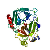
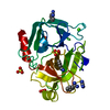
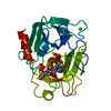
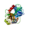
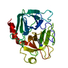
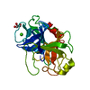
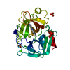
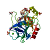
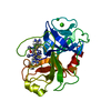
| Entry | Database: PDB / ID: 1j14 | ||||||
|---|---|---|---|---|---|---|---|
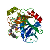
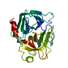
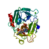
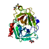
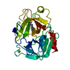
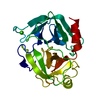
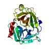
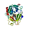
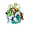
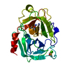
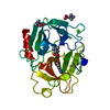
| Title | BENZAMIDINE IN COMPLEX WITH RAT TRYPSIN MUTANT X99RT | ||||||
 Components Components | trypsin II, anionic | ||||||
 Keywords Keywords | HYDROLASE / SERINE PROTEASE / SERINE PROTEINASE | ||||||
| Function / homology |  Function and homology information Function and homology informationAntimicrobial peptides / Alpha-defensins / Activation of Matrix Metalloproteinases / Neutrophil degranulation / collagen catabolic process / trypsin / digestion / response to nutrient / serine-type endopeptidase activity / calcium ion binding ...Antimicrobial peptides / Alpha-defensins / Activation of Matrix Metalloproteinases / Neutrophil degranulation / collagen catabolic process / trypsin / digestion / response to nutrient / serine-type endopeptidase activity / calcium ion binding / proteolysis / : / extracellular region Similarity search - Function | ||||||
| Biological species |  | ||||||
| Method |  X-RAY DIFFRACTION / Resolution: 2.4 Å X-RAY DIFFRACTION / Resolution: 2.4 Å | ||||||
 Authors Authors | Stubbs, M.T. | ||||||
 Citation Citation | Journal: J.MOL.BIOL. / Year: 2003 Title: Reconstructing the Binding Site of Factor Xa in Trypsin Reveals Ligand-induced Structural Plasticity Authors: Reyda, S. / Sohn, C. / Klebe, G. / Rall, K. / Ullmann, D. / Jakubke, H.D. / Stubbs, M.T. #1: Journal: Chembiochem / Year: 2002Title: pH-dependent binding modes observed in trypsin crystals: lessons for structure-based drug design Authors: Stubbs, M.T. / Reyda, S. / Dullweber, F. / Moeller, M. / Klebe, G. / Dorsch, D. / Mederski, W.W.K.R. / Wurziger, H. #2: Journal: J.MED.CHEM. / Year: 1998Title: Structural and functional analyses of benzamidine-based inhibitors in complex with trypsin: implications for the inhibition of factor Xa, tPA, and urokinas Authors: Renatus, M. / Bode, W. / Huber, R. / Stuerzebecher, J. / Stubbs, M.T. #3: Journal: Curr.Pharm.Des. / Year: 1996Title: Structural Aspects of Factor Xa Inhibition Authors: Stubbs, M.T. #4: Journal: FEBS LETT. / Year: 1995Title: Crystal structures of factor Xa specific inhibitors in complex with trypsin: structural grounds for inhibition of factor Xa and selectivity against thrombin Authors: Stubbs, M.T. / Huber, R. / Bode, W. | ||||||
| History |
|
- Structure visualization
| Structure viewer | Molecule: MolmilJmol/JSmol |
|---|
- Downloads & links
-Download
| PDBx/mmCIF format | 1j14.cif.gz | 56.5 KB | Display | PDBx/mmCIF format |
|---|---|---|---|---|
| PDB format | pdb1j14.ent.gz | 40.2 KB | Display | PDB format |
| PDBx/mmJSON format | 1j14.json.gz | Tree view | PDBx/mmJSON format | |
| Others |  Other downloads Other downloads |
-Validation report
| Arichive directory | https://data.pdbj.org/pub/pdb/validation_reports/j1/1j14ftp://data.pdbj.org/pub/pdb/validation_reports/j1/1j14 | HTTPS FTP |
|---|
-Related structure data


-Links
 PDBj
PDBj


- Assembly
| Deposited unit | 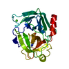
| ||||||||
|---|---|---|---|---|---|---|---|---|---|
| 1 |
| ||||||||
| Unit cell |
|
-Components
| #1: Protein | Mass: 23864.787 Da / Num. of mol.: 1 / Mutation: K97E, L99Y Source method: isolated from a genetically manipulated source Source: (gene. exp.)  |
|---|---|
| #2: Chemical | ChemComp-CA /   Mass: 40.078 Da / Num. of mol.: 1 / Source method: obtained synthetically / Formula: Ca Mass: 40.078 Da / Num. of mol.: 1 / Source method: obtained synthetically / Formula: Ca |
| #3: Chemical | ChemComp-SO4 /   Mass: 96.063 Da / Num. of mol.: 1 / Source method: obtained synthetically / Formula: SO4 Mass: 96.063 Da / Num. of mol.: 1 / Source method: obtained synthetically / Formula: SO4 |
| #4: Chemical | ChemComp-BEN /   Mass: 120.152 Da / Num. of mol.: 1 / Source method: obtained synthetically / Formula: C7H8N2 Mass: 120.152 Da / Num. of mol.: 1 / Source method: obtained synthetically / Formula: C7H8N2 |
| #5: Water | ChemComp-HOH /  Mass: 18.015 Da / Num. of mol.: 64 / Source method: isolated from a natural source / Formula: H2O Mass: 18.015 Da / Num. of mol.: 64 / Source method: isolated from a natural source / Formula: H2O |
| Has protein modification | Y |
-Experimental details
-Experiment
| Experiment | Method: X-RAY DIFFRACTION / Number of used crystals: 1 |
|---|
- Sample preparation
| Crystal | Density Matthews: 3.29 Å3/Da / Density % sol: 62.27 % |
|---|---|
| Crystal grow | pH: 7 / Details: pH 7.0 |
-Data collection
| Diffraction | Mean temperature: 287 K |
|---|---|
| Diffraction source | Source: ROTATING ANODE / Type: RIGAKU RU300 / Wavelength: 1.5418 Å |
| Detector | Type: RIGAKU RAXIS IV / Detector: IMAGE PLATE / Date: Jun 4, 1998 |
| Radiation | Monochromator: NI FILTER / Protocol: SINGLE WAVELENGTH / Monochromatic (M) / Laue (L): M / Scattering type: x-ray |
| Radiation wavelength | Wavelength: 1.5418 Å / Relative weight: 1 |
| Reflection | Resolution: 2.4→50.84 Å / Num. obs: 12713 |
- Processing
| Software |
| ||||||||||||
|---|---|---|---|---|---|---|---|---|---|---|---|---|---|
| Refinement | Resolution: 2.4→10 Å / σ(F): 0 /
| ||||||||||||
| Refinement step | Cycle: LAST / Resolution: 2.4→10 Å
|
