 Movie
Movie Controller


+ Open data

- Basic information
| Entry | Database: PDB / ID: 1j02 | ||||||
|---|---|---|---|---|---|---|---|
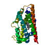
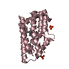
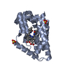
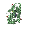
| Title | Crystal Structure of Rat Heme Oxygenase-1-Heme Bound to NO | ||||||
 Components Components | HEME OXYGENASE 1 | ||||||
 Keywords Keywords | OXIDOREDUCTASE / alpha helix / O2-analog bound form | ||||||
| Function / homology |  Function and homology information Function and homology informationRegulation of HMOX1 expression and activity / arachidonate omega-hydroxylase activity / Iron uptake and transport / Heme degradation / Cytoprotection by HMOX1 / negative regulation of mast cell degranulation / response to arachidonate / response to 3-methylcholanthrene / negative regulation of muscle cell apoptotic process / heme metabolic process ...Regulation of HMOX1 expression and activity / arachidonate omega-hydroxylase activity / Iron uptake and transport / Heme degradation / Cytoprotection by HMOX1 / negative regulation of mast cell degranulation / response to arachidonate / response to 3-methylcholanthrene / negative regulation of muscle cell apoptotic process / heme metabolic process / heme oxygenase (biliverdin-producing) / heme oxidation / heme oxygenase (decyclizing) activity / Insertion of tail-anchored proteins into the endoplasmic reticulum membrane / wound healing involved in inflammatory response / cellular response to arsenic-containing substance / cellular response to cisplatin / positive regulation of blood vessel endothelial cell proliferation involved in sprouting angiogenesis / negative regulation of epithelial cell apoptotic process / heme catabolic process / cellular response to nutrient / negative regulation of viral life cycle / negative regulation of mast cell cytokine production / D-type glycerophospholipase activity / positive regulation of epithelial cell apoptotic process / positive regulation of cell migration involved in sprouting angiogenesis / small GTPase-mediated signal transduction / negative regulation of macroautophagy / erythrocyte homeostasis / cellular response to cadmium ion / negative regulation of ferroptosis / negative regulation of vascular associated smooth muscle cell proliferation / positive regulation of macroautophagy / host-mediated suppression of viral transcription / negative regulation of extrinsic apoptotic signaling pathway via death domain receptors / phospholipid metabolic process / negative regulation of cytokine production involved in inflammatory response / liver regeneration / positive regulation of smooth muscle cell proliferation / erythrocyte differentiation / response to nicotine / negative regulation of smooth muscle cell proliferation / response to hydrogen peroxide / multicellular organismal-level iron ion homeostasis / caveola / response to estrogen / regulation of blood pressure / intrinsic apoptotic signaling pathway in response to DNA damage / positive regulation of angiogenesis / cellular response to heat / response to oxidative stress / angiogenesis / intracellular iron ion homeostasis / negative regulation of neuron apoptotic process / response to hypoxia / intracellular signal transduction / response to xenobiotic stimulus / negative regulation of cell population proliferation / heme binding / regulation of transcription by RNA polymerase II / endoplasmic reticulum membrane / perinuclear region of cytoplasm / structural molecule activity / enzyme binding / endoplasmic reticulum / protein homodimerization activity / metal ion binding / identical protein binding / nucleus / cytosol Similarity search - Function | ||||||
| Biological species |  | ||||||
| Method |  X-RAY DIFFRACTION / SYNCHROTRON / MOLECULAR REPLACEMENT / Resolution: 1.7 Å X-RAY DIFFRACTION / SYNCHROTRON / MOLECULAR REPLACEMENT / Resolution: 1.7 Å | ||||||
 Authors Authors | Sugishima, M. / Fukuyama, K. | ||||||
 Citation Citation | Journal: Biochemistry / Year: 2003 Title: Crystal Structures of Ferrous and CO-, CN(-)-, and NO-Bound Forms of Rat Heme Oxygenase-1 (HO-1) in Complex with Heme: Structural Implications for Discrimination between CO and O(2) in HO-1. Authors: Sugishima, M. / Sakamoto, H. / Noguchi, M. / Fukuyama, K. | ||||||
| History |
|
- Structure visualization
| Structure viewer | Molecule: MolmilJmol/JSmol |
|---|
- Downloads & links
-Download
| PDBx/mmCIF format | 1j02.cif.gz | 64.4 KB | Display | PDBx/mmCIF format |
|---|---|---|---|---|
| PDB format | pdb1j02.ent.gz | 45.8 KB | Display | PDB format |
| PDBx/mmJSON format | 1j02.json.gz | Tree view | PDBx/mmJSON format | |
| Others |  Other downloads Other downloads |
-Validation report
| Arichive directory | https://data.pdbj.org/pub/pdb/validation_reports/j0/1j02ftp://data.pdbj.org/pub/pdb/validation_reports/j0/1j02 | HTTPS FTP |
|---|
-Related structure data
| Related structure data |  1ix3C  1ix4C  1ubbC  1ivjS S: Starting model for refinement C: citing same article ( |
|---|---|
| Similar structure data |


-Links
 PDBj
PDBj


- Assembly
| Deposited unit | 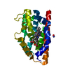
| ||||||||
|---|---|---|---|---|---|---|---|---|---|
| 1 |
| ||||||||
| Unit cell |
|
-Components
| #1: Protein | Mass: 30612.496 Da / Num. of mol.: 1 / Fragment: C-terminal truncated fragment Source method: isolated from a genetically manipulated source Source: (gene. exp.)  References: UniProt: P06762, heme oxygenase (biliverdin-producing) | ||
|---|---|---|---|
| #2: Chemical | ChemComp-HEM /   Mass: 616.487 Da / Num. of mol.: 1 / Source method: obtained synthetically / Formula: C34H32FeN4O4 Mass: 616.487 Da / Num. of mol.: 1 / Source method: obtained synthetically / Formula: C34H32FeN4O4 | ||
| #3: Chemical |   Mass: 30.006 Da / Num. of mol.: 2 / Source method: obtained synthetically / Formula: NO Mass: 30.006 Da / Num. of mol.: 2 / Source method: obtained synthetically / Formula: NO#4: Water | ChemComp-HOH / |  Mass: 18.015 Da / Num. of mol.: 167 / Source method: isolated from a natural source / Formula: H2O Mass: 18.015 Da / Num. of mol.: 167 / Source method: isolated from a natural source / Formula: H2O |
-Experimental details
-Experiment
| Experiment | Method: X-RAY DIFFRACTION / Number of used crystals: 1 |
|---|
- Sample preparation
| Crystal | Density Matthews: 2.67 Å3/Da / Density % sol: 53.59 % | ||||||||||||||||||||||||||||||
|---|---|---|---|---|---|---|---|---|---|---|---|---|---|---|---|---|---|---|---|---|---|---|---|---|---|---|---|---|---|---|---|
| Crystal grow | Temperature: 293 K / Method: vapor diffusion, hanging drop / pH: 7 Details: sodium formate, potassium phosphate, sodium azide, pH 7.0, VAPOR DIFFUSION, HANGING DROP, temperature 293K | ||||||||||||||||||||||||||||||
| Crystal grow | *PLUS Method: vapor diffusion, hanging drop / Details: Sugishima, M., (2002) J.Biol.Chem., 277, 45086. | ||||||||||||||||||||||||||||||
| Components of the solutions | *PLUS
|
-Data collection
| Diffraction | Mean temperature: 100 K |
|---|---|
| Diffraction source | Source: SYNCHROTRON / Site: SPring-8  / Beamline: BL41XU / Wavelength: 1 Å / Beamline: BL41XU / Wavelength: 1 Å |
| Detector | Type: MARRESEARCH / Detector: CCD / Date: Oct 8, 2002 |
| Radiation | Monochromator: Si(111) double monochrometer / Protocol: SINGLE WAVELENGTH / Monochromatic (M) / Laue (L): M / Scattering type: x-ray |
| Radiation wavelength | Wavelength: 1 Å / Relative weight: 1 |
| Reflection | Resolution: 1.7→50 Å / Num. all: 33605 / Num. obs: 32251 / % possible obs: 95.8 % / Observed criterion σ(F): 0 / Observed criterion σ(I): 0 / Redundancy: 9.5 % / Rmerge(I) obs: 0.052 / Rsym value: 0.052 / Net I/σ(I): 18.1 |
| Reflection shell | Resolution: 1.7→1.76 Å / Rmerge(I) obs: 0.346 / Num. unique all: 2919 / Rsym value: 0.346 / % possible all: 87.9 |
| Reflection | *PLUS % possible obs: 99.8 % |
| Reflection shell | *PLUS % possible obs: 99.5 % |
- Processing
| Software |
| ||||||||||||||||||||||||||||||||||||
|---|---|---|---|---|---|---|---|---|---|---|---|---|---|---|---|---|---|---|---|---|---|---|---|---|---|---|---|---|---|---|---|---|---|---|---|---|---|
| Refinement | Method to determine structure: MOLECULAR REPLACEMENT Starting model: PDB ENTRY 1IVJ Resolution: 1.7→50 Å / Cross valid method: THROUGHOUT / σ(F): 0 / Stereochemistry target values: Engh & Huber
| ||||||||||||||||||||||||||||||||||||
| Solvent computation | Solvent model: FLAT MODEL / Bsol: 53.2 Å2 / ksol: 0.382 e/Å3 | ||||||||||||||||||||||||||||||||||||
| Displacement parameters |
| ||||||||||||||||||||||||||||||||||||
| Refine analyze |
| ||||||||||||||||||||||||||||||||||||
| Refinement step | Cycle: LAST / Resolution: 1.7→50 Å
| ||||||||||||||||||||||||||||||||||||
| Refine LS restraints |
| ||||||||||||||||||||||||||||||||||||
| LS refinement shell | Resolution: 1.7→1.76 Å
| ||||||||||||||||||||||||||||||||||||
| Xplor file |
| ||||||||||||||||||||||||||||||||||||
| Software | *PLUS Version: 1.1 / Classification: refinement | ||||||||||||||||||||||||||||||||||||
| Refinement | *PLUS % reflection Rfree: 5 % | ||||||||||||||||||||||||||||||||||||
| Solvent computation | *PLUS | ||||||||||||||||||||||||||||||||||||
| Displacement parameters | *PLUS | ||||||||||||||||||||||||||||||||||||
| Refine LS restraints | *PLUS
|
