 Movie
Movie Controller

[English] 日本語
 Yorodumi
- PDB-1iz6: Crystal Structure of Translation Initiation Factor 5A from Pyroco... -
+ Open data

- Basic information
| Entry | Database: PDB / ID: 1iz6 | ||||||
|---|---|---|---|---|---|---|---|
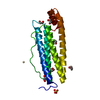
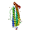
| Title | Crystal Structure of Translation Initiation Factor 5A from Pyrococcus Horikoshii | ||||||
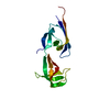
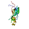
 Components Components | Initiation Factor 5A | ||||||
 Keywords Keywords | BIOSYNTHETIC PROTEIN / SH3-like barrel / OB fold | ||||||
| Function / homology |  Function and homology information Function and homology informationpositive regulation of translational termination / positive regulation of translational elongation / translation elongation factor activity / translation initiation factor activity / ribosome binding / RNA binding / cytoplasm Similarity search - Function | ||||||
| Biological species |   Pyrococcus horikoshii (archaea) Pyrococcus horikoshii (archaea) | ||||||
| Method |  X-RAY DIFFRACTION / SYNCHROTRON / MOLECULAR REPLACEMENT / Resolution: 2 Å X-RAY DIFFRACTION / SYNCHROTRON / MOLECULAR REPLACEMENT / Resolution: 2 Å | ||||||
 Authors Authors | Yao, M. / Ohsawa, A. / Kikukawa, S. / Tanaka, I. / Kimura, M. | ||||||
 Citation Citation | Journal: J.BIOCHEM.(TOKYO) / Year: 2003 Title: Crystal Structure of Hyperthermophilic Archaeal Initiation Factor 5A: A Homologue of Eukaryotic Initiation Factor 5A (eIF-5A) Authors: Yao, M. / Ohsawa, A. / Kikukawa, S. / Tanaka, I. / Kimura, M. | ||||||
| History |
|
- Structure visualization
| Structure viewer | Molecule: MolmilJmol/JSmol |
|---|
- Downloads & links
-Download
| PDBx/mmCIF format | 1iz6.cif.gz | 94.3 KB | Display | PDBx/mmCIF format |
|---|---|---|---|---|
| PDB format | pdb1iz6.ent.gz | 72.9 KB | Display | PDB format |
| PDBx/mmJSON format | 1iz6.json.gz | Tree view | PDBx/mmJSON format | |
| Others |  Other downloads Other downloads |
-Validation report
| Arichive directory | https://data.pdbj.org/pub/pdb/validation_reports/iz/1iz6ftp://data.pdbj.org/pub/pdb/validation_reports/iz/1iz6 | HTTPS FTP |
|---|
-Related structure data
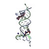
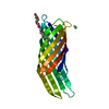
| Related structure data | 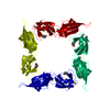 1bkbS S: Starting model for refinement |
|---|---|
| Similar structure data |


-Links
 PDBj
PDBj- Assembly
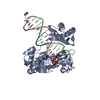
| Deposited unit | 
| ||||||||
|---|---|---|---|---|---|---|---|---|---|
| 1 | 
| ||||||||
| 2 | 
| ||||||||
| 3 | 
| ||||||||
| Unit cell |
|
-Components
| #1: Protein | Mass: 15305.637 Da / Num. of mol.: 3 Source method: isolated from a genetically manipulated source Source: (gene. exp.) Pyrococcus horikoshii (archaea) / Strain: OT3 / Gene: PH1381 / Plasmid: pET-22b / Species (production host): Escherichia coli / Production host:  #2: Water | ChemComp-HOH / |  Mass: 18.015 Da / Num. of mol.: 279 / Source method: isolated from a natural source / Formula: H2O Mass: 18.015 Da / Num. of mol.: 279 / Source method: isolated from a natural source / Formula: H2O |
|---|
-Experimental details
-Experiment
| Experiment | Method: X-RAY DIFFRACTION / Number of used crystals: 1 |
|---|
- Sample preparation
| Crystal | Density Matthews: 2.16 Å3/Da / Density % sol: 43.1 % | ||||||||||||||||||||||||||||||||||||
|---|---|---|---|---|---|---|---|---|---|---|---|---|---|---|---|---|---|---|---|---|---|---|---|---|---|---|---|---|---|---|---|---|---|---|---|---|---|
| Crystal grow | Temperature: 291 K / Method: vapor diffusion, hanging drop / pH: 7.5 Details: PEG 4000, isopropanol, Hepers, pH 7.5, VAPOR DIFFUSION, HANGING DROP, temperature 291K | ||||||||||||||||||||||||||||||||||||
| Crystal grow | *PLUS Temperature: 18 ℃ | ||||||||||||||||||||||||||||||||||||
| Components of the solutions | *PLUS
|
-Data collection
| Diffraction | Mean temperature: 100 K |
|---|---|
| Diffraction source | Source: SYNCHROTRON / Site: SPring-8  / Beamline: BL41XU / Wavelength: 0.9 Å / Beamline: BL41XU / Wavelength: 0.9 Å |
| Detector | Type: MARRESEARCH / Detector: CCD / Date: Sep 20, 2001 |
| Radiation | Monochromator: MIRROR / Protocol: SINGLE WAVELENGTH / Monochromatic (M) / Laue (L): M / Scattering type: x-ray |
| Radiation wavelength | Wavelength: 0.9 Å / Relative weight: 1 |
| Reflection | Resolution: 2→20 Å / Num. obs: 25949 / % possible obs: 100 % / Observed criterion σ(I): 3 / Redundancy: 5.7 % / Biso Wilson estimate: 42.5 Å2 / Rmerge(I) obs: 0.081 / Rsym value: 0.074 / Net I/σ(I): 8.5 |
| Reflection shell | Resolution: 2→2.11 Å / Redundancy: 5.7 % / Rmerge(I) obs: 0.349 / Mean I/σ(I) obs: 2.4 / Num. unique all: 3842 / Rsym value: 0.317 / % possible all: 99.9 |
| Reflection | *PLUS Lowest resolution: 100 Å / % possible obs: 100 % / Num. measured all: 148604 |
| Reflection shell | *PLUS % possible obs: 99.9 % |
- Processing
| Software |
| |||||||||||||||||||||||||||
|---|---|---|---|---|---|---|---|---|---|---|---|---|---|---|---|---|---|---|---|---|---|---|---|---|---|---|---|---|
| Refinement | Method to determine structure: MOLECULAR REPLACEMENT Starting model: PDB ENTRY 1BKB Resolution: 2→10 Å / Isotropic thermal model: isotropic / Cross valid method: THROUGHOUT / σ(F): 0 / σ(I): 0 / Stereochemistry target values: Engh & Huber
| |||||||||||||||||||||||||||
| Solvent computation | Solvent model: throughout / Bsol: 77.45 Å2 / ksol: 0.4885 e/Å3 | |||||||||||||||||||||||||||
| Displacement parameters | Biso mean: 28.8 Å2
| |||||||||||||||||||||||||||
| Refine analyze |
| |||||||||||||||||||||||||||
| Refinement step | Cycle: LAST / Resolution: 2→10 Å
| |||||||||||||||||||||||||||
| Refine LS restraints |
| |||||||||||||||||||||||||||
| LS refinement shell | Refine-ID: X-RAY DIFFRACTION / Total num. of bins used: 10 / % reflection obs: 100 %
| |||||||||||||||||||||||||||
| Xplor file | Serial no: 1 / Param file: protein_rep.param / Topol file: protein.top | |||||||||||||||||||||||||||
| Refinement | *PLUS Num. reflection obs: 25746 / % reflection Rfree: 10 % | |||||||||||||||||||||||||||
| Solvent computation | *PLUS | |||||||||||||||||||||||||||
| Displacement parameters | *PLUS | |||||||||||||||||||||||||||
| Refine LS restraints | *PLUS
|
