 Movie
Movie Controller


+ Open data

- Basic information
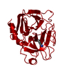
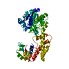
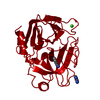
| Entry | Database: PDB / ID: 1iyb | ||||||
|---|---|---|---|---|---|---|---|
| Title | Crystal Structure of the Nicotiana glutinosa Ribonuclease NW | ||||||
 Components Components | Ribonuclease | ||||||
 Keywords Keywords | HYDROLASE / ribonuclease | ||||||
| Function / homology |  Function and homology information Function and homology informationribonuclease T2 activity / RNA catabolic process / hydrolase activity / RNA binding / extracellular region Similarity search - Function | ||||||
| Biological species |  | ||||||
| Method |  X-RAY DIFFRACTION / SYNCHROTRON / MOLECULAR REPLACEMENT / Resolution: 1.5 Å X-RAY DIFFRACTION / SYNCHROTRON / MOLECULAR REPLACEMENT / Resolution: 1.5 Å | ||||||
 Authors Authors | Kawano, S. / Kakuta, Y. / Kimura, M. | ||||||
 Citation Citation | Journal: Biochemistry / Year: 2002 Title: Guanine binding site of the Nicotiana glutinosa ribonuclease NW revealed by X-ray crystallography Authors: Kawano, S. / Kakuta, Y. / Kimura, M. | ||||||
| History |
|
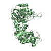
- Structure visualization
| Structure viewer | Molecule: MolmilJmol/JSmol |
|---|
- Downloads & links
-Download
| PDBx/mmCIF format | 1iyb.cif.gz | 102.7 KB | Display | PDBx/mmCIF format |
|---|---|---|---|---|
| PDB format | pdb1iyb.ent.gz | 78.1 KB | Display | PDB format |
| PDBx/mmJSON format | 1iyb.json.gz | Tree view | PDBx/mmJSON format | |
| Others |  Other downloads Other downloads |
-Validation report
| Summary document | 1iyb_validation.pdf.gz | 1.1 MB | Display | wwPDB validaton report |
|---|---|---|---|---|
| Full document | 1iyb_full_validation.pdf.gz | 1.1 MB | Display | |
| Data in XML | 1iyb_validation.xml.gz | 21.7 KB | Display | |
| Data in CIF | 1iyb_validation.cif.gz | 32 KB | Display | |
| Arichive directory | https://data.pdbj.org/pub/pdb/validation_reports/iy/1iybftp://data.pdbj.org/pub/pdb/validation_reports/iy/1iyb | HTTPS FTP |
-Related structure data
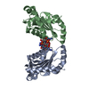
| Related structure data |  1dixS S: Starting model for refinement |
|---|---|
| Similar structure data |


-Links
 PDBj
PDBj

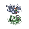
- Assembly
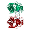
| Deposited unit | 
| ||||||||
|---|---|---|---|---|---|---|---|---|---|
| 1 |
| ||||||||
| Unit cell |
|
-Components
| #1: Protein | Mass: 23126.467 Da / Num. of mol.: 2 Source method: isolated from a genetically manipulated source Source: (gene. exp.)  Pichia pastoris (fungus) Pichia pastoris (fungus)References: UniProt: Q7XZV5, Hydrolases; Acting on ester bonds #2: Chemical |   Mass: 363.221 Da / Num. of mol.: 2 / Source method: obtained synthetically / Formula: C10H14N5O8P Mass: 363.221 Da / Num. of mol.: 2 / Source method: obtained synthetically / Formula: C10H14N5O8P#3: Water | ChemComp-HOH / |  Mass: 18.015 Da / Num. of mol.: 410 / Source method: isolated from a natural source / Formula: H2O Mass: 18.015 Da / Num. of mol.: 410 / Source method: isolated from a natural source / Formula: H2OHas protein modification | Y | |
|---|
-Experimental details
-Experiment
| Experiment | Method: X-RAY DIFFRACTION / Number of used crystals: 1 |
|---|
- Sample preparation
| Crystal | Density Matthews: 2.24 Å3/Da / Density % sol: 45.06 % | ||||||||||||||||||||||||
|---|---|---|---|---|---|---|---|---|---|---|---|---|---|---|---|---|---|---|---|---|---|---|---|---|---|
| Crystal grow | Temperature: 291.15 K / Method: vapor diffusion, hanging drop / pH: 8.5 Details: ammonium dihydrogen, Tris-HCl, pH 8.5, VAPOR DIFFUSION, HANGING DROP, temperature 291.15K | ||||||||||||||||||||||||
| Crystal grow | *PLUS Temperature: 18 ℃ / pH: 7 / Method: vapor diffusion, hanging drop | ||||||||||||||||||||||||
| Components of the solutions | *PLUS
|
-Data collection
| Diffraction | Mean temperature: 100 K |
|---|---|
| Diffraction source | Source: SYNCHROTRON / Site: SPring-8  / Beamline: BL44XU / Wavelength: 0.9 Å / Beamline: BL44XU / Wavelength: 0.9 Å |
| Detector | Type: OXFORD PX210 / Detector: CCD |
| Radiation | Monochromator: Si 111 / Protocol: SINGLE WAVELENGTH / Monochromatic (M) / Laue (L): M / Scattering type: x-ray |
| Radiation wavelength | Wavelength: 0.9 Å / Relative weight: 1 |
| Reflection | Resolution: 1.5→37.43 Å / Num. obs: 62367 / % possible obs: 93.2 % / Observed criterion σ(F): 1 / Observed criterion σ(I): 1 / Redundancy: 5.6 % / Biso Wilson estimate: 14.6 Å2 / Rmerge(I) obs: 0.052 / Net I/σ(I): 28.6 |
| Reflection shell | Resolution: 1.5→1.55 Å / Redundancy: 1.7 % / Rmerge(I) obs: 0.201 / Mean I/σ(I) obs: 2.7 / Num. unique all: 3581 / % possible all: 53.8 |
| Reflection | *PLUS Highest resolution: 1.5 Å / Lowest resolution: 37.4 Å |
| Reflection shell | *PLUS % possible obs: 53.8 % / Mean I/σ(I) obs: 2 |
- Processing
| Software |
| ||||||||||||||||||||||||||||||||||||
|---|---|---|---|---|---|---|---|---|---|---|---|---|---|---|---|---|---|---|---|---|---|---|---|---|---|---|---|---|---|---|---|---|---|---|---|---|---|
| Refinement | Method to determine structure: MOLECULAR REPLACEMENT Starting model: PDB ENTRY 1DIX Resolution: 1.5→37.41 Å / Rfactor Rfree error: 0.004 / Data cutoff high absF: 315208.91 / Data cutoff low absF: 0 / Isotropic thermal model: RESTRAINED / Cross valid method: THROUGHOUT / σ(F): 0 / σ(I): 0
| ||||||||||||||||||||||||||||||||||||
| Solvent computation | Solvent model: FLAT MODEL / Bsol: 39.5283 Å2 / ksol: 0.386094 e/Å3 | ||||||||||||||||||||||||||||||||||||
| Displacement parameters | Biso mean: 13 Å2
| ||||||||||||||||||||||||||||||||||||
| Refine analyze |
| ||||||||||||||||||||||||||||||||||||
| Refinement step | Cycle: LAST / Resolution: 1.5→37.41 Å
| ||||||||||||||||||||||||||||||||||||
| Refine LS restraints |
| ||||||||||||||||||||||||||||||||||||
| LS refinement shell | Resolution: 1.5→1.59 Å / Rfactor Rfree error: 0.013 / Total num. of bins used: 6
| ||||||||||||||||||||||||||||||||||||
| Xplor file |
| ||||||||||||||||||||||||||||||||||||
| Refinement | *PLUS Highest resolution: 1.5 Å / Lowest resolution: 37.4 Å / % reflection Rfree: 5 % / Rfactor Rfree: 0.23 | ||||||||||||||||||||||||||||||||||||
| Solvent computation | *PLUS | ||||||||||||||||||||||||||||||||||||
| Displacement parameters | *PLUS | ||||||||||||||||||||||||||||||||||||
| Refine LS restraints | *PLUS
|
