 Movie
Movie Controller

[English] 日本語
 Yorodumi
- PDB-1ixx: CRYSTAL STRUCTURE OF COAGULATION FACTORS IX/X-BINDING PROTEIN (IX... -
+ Open data

- Basic information
| Entry | Database: PDB / ID: 1ixx | ||||||
|---|---|---|---|---|---|---|---|
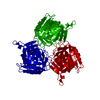
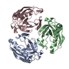
| Title | CRYSTAL STRUCTURE OF COAGULATION FACTORS IX/X-BINDING PROTEIN (IX/X-BP) FROM VENOM OF HABU SNAKE WITH A HETERODIMER OF C-TYPE LECTIN DOMAINS | ||||||
 Components Components | (COAGULATION FACTORS IX/X-BINDING PROTEIN) x 2 | ||||||
 Keywords Keywords | COAGULATION FACTOR BINDING / C-TYPE LECTIN / GLA-DOMAIN BINDING / C-TYPE CRD MOTIF / LOOP EXCHANGED DIMER | ||||||
| Function / homology |  Function and homology information Function and homology informationsignaling receptor activity / toxin activity / calcium ion binding / extracellular region / metal ion binding Similarity search - Function | ||||||
| Biological species |  Trimeresurus flavoviridis (habu) Trimeresurus flavoviridis (habu) | ||||||
| Method |  X-RAY DIFFRACTION / SYNCHROTRON / SIRAS / Resolution: 2.5 Å X-RAY DIFFRACTION / SYNCHROTRON / SIRAS / Resolution: 2.5 Å | ||||||
 Authors Authors | Mizuno, H. / Fujimoto, Z. / Koizumi, M. / Kano, H. | ||||||
 Citation Citation | Journal: Nat.Struct.Biol. / Year: 1997 Title: Structure of coagulation factors IX/X-binding protein, a heterodimer of C-type lectin domains. Authors: Mizuno, H. / Fujimoto, Z. / Koizumi, M. / Kano, H. / Atoda, H. / Morita, T. | ||||||
| History |
|
- Structure visualization
| Structure viewer | Molecule: MolmilJmol/JSmol |
|---|
- Downloads & links
-Download
| PDBx/mmCIF format | 1ixx.cif.gz | 161.2 KB | Display | PDBx/mmCIF format |
|---|---|---|---|---|
| PDB format | pdb1ixx.ent.gz | 135 KB | Display | PDB format |
| PDBx/mmJSON format | 1ixx.json.gz | Tree view | PDBx/mmJSON format | |
| Others |  Other downloads Other downloads |
-Validation report
| Summary document | 1ixx_validation.pdf.gz | 395.5 KB | Display | wwPDB validaton report |
|---|---|---|---|---|
| Full document | 1ixx_full_validation.pdf.gz | 409.8 KB | Display | |
| Data in XML | 1ixx_validation.xml.gz | 16.8 KB | Display | |
| Data in CIF | 1ixx_validation.cif.gz | 26 KB | Display | |
| Arichive directory | https://data.pdbj.org/pub/pdb/validation_reports/ix/1ixxftp://data.pdbj.org/pub/pdb/validation_reports/ix/1ixx | HTTPS FTP |
-Related structure data
| Similar structure data |
|---|

-Links
 PDBj
PDBj


- Assembly
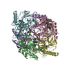
| Deposited unit | 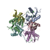
| ||||||||||||
|---|---|---|---|---|---|---|---|---|---|---|---|---|---|
| 1 |
| ||||||||||||
| 2 | 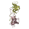
| ||||||||||||
| 3 | 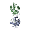
| ||||||||||||
| 4 | 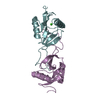
| ||||||||||||
| Unit cell |
| ||||||||||||
| Noncrystallographic symmetry (NCS) | NCS oper:
| ||||||||||||
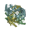
| Details | THE ASYMMETRIC UNIT CONTAINS THREE DIMERS ARRANGED AROUND A PSEUDO TRIAD. |
-Components
| #1: Protein | Mass: 14845.512 Da / Num. of mol.: 3 / Fragment: C-TYPE LECTIN DOMAIN / Source method: isolated from a natural source / Source: (natural) Trimeresurus flavoviridis (habu) / Secretion: VENOM / References: UniProt: P23806#2: Protein | Mass: 14455.071 Da / Num. of mol.: 3 / Fragment: C-TYPE LECTIN DOMAIN / Source method: isolated from a natural source / Source: (natural) Trimeresurus flavoviridis (habu) / Secretion: VENOM / References: UniProt: P23807#3: Chemical | ChemComp-CA /   Mass: 40.078 Da / Num. of mol.: 6 / Source method: obtained synthetically / Formula: Ca Mass: 40.078 Da / Num. of mol.: 6 / Source method: obtained synthetically / Formula: Ca#4: Water | ChemComp-HOH / |  Mass: 18.015 Da / Num. of mol.: 146 / Source method: isolated from a natural source / Formula: H2O Mass: 18.015 Da / Num. of mol.: 146 / Source method: isolated from a natural source / Formula: H2OCompound details | IX/X-BP IS A DISULFIDE-LINKED HETERODIMER PROTEIN CONSISTING OF HOMOLOGOUS SUBUNITS. EACH SUBUNIT ...IX/X-BP IS A DISULFIDE-LINKED HETERODIME | |
|---|
-Experimental details
-Experiment
| Experiment | Method: X-RAY DIFFRACTION / Number of used crystals: 2 |
|---|
- Sample preparation
| Crystal | Density Matthews: 3 Å3/Da / Density % sol: 59 % | ||||||||||||||||||||||||||||||||||||
|---|---|---|---|---|---|---|---|---|---|---|---|---|---|---|---|---|---|---|---|---|---|---|---|---|---|---|---|---|---|---|---|---|---|---|---|---|---|
| Crystal grow | pH: 7.8 Details: 60% SATURATED AMMONIUM SULFATE, 20 MM TRIS-HCL, 3 MM CACL2, PH 7.8 | ||||||||||||||||||||||||||||||||||||
| Crystal | *PLUS Density % sol: 70 % | ||||||||||||||||||||||||||||||||||||
| Crystal grow | *PLUS Method: vapor diffusion, hanging drop / Details: Mizuno, H., (1991) J. Mol. Biol., 220, 225. | ||||||||||||||||||||||||||||||||||||
| Components of the solutions | *PLUS
|
-Data collection
| Diffraction | Mean temperature: 288 K |
|---|---|
| Diffraction source | Source: SYNCHROTRON / Site: Photon Factory  / Beamline: BL-6A / Wavelength: 1 / Beamline: BL-6A / Wavelength: 1 |
| Detector | Type: RIGAKU / Detector: IMAGE PLATE / Date: May 28, 1996 |
| Radiation | Monochromatic (M) / Laue (L): M / Scattering type: x-ray |
| Radiation wavelength | Wavelength: 1 Å / Relative weight: 1 |
| Reflection | Resolution: 2.5→50 Å / Num. obs: 29883 / % possible obs: 87 % / Observed criterion σ(I): 1 / Redundancy: 3.8 % / Biso Wilson estimate: 30.6 Å2 / Rmerge(I) obs: 0.053 / Net I/σ(I): 39 |
| Reflection shell | Resolution: 2.5→2.61 Å / Rmerge(I) obs: 0.229 / Mean I/σ(I) obs: 6 / % possible all: 75 |
- Processing
| Software |
| ||||||||||||||||||||||||||||||||||||||||||||||||||||||||||||||||||||||||||||||||
|---|---|---|---|---|---|---|---|---|---|---|---|---|---|---|---|---|---|---|---|---|---|---|---|---|---|---|---|---|---|---|---|---|---|---|---|---|---|---|---|---|---|---|---|---|---|---|---|---|---|---|---|---|---|---|---|---|---|---|---|---|---|---|---|---|---|---|---|---|---|---|---|---|---|---|---|---|---|---|---|---|---|
| Refinement | Method to determine structure: SIRAS / Resolution: 2.5→6 Å / Rfactor Rfree error: 0.006 / Data cutoff high absF: 2000000 / Data cutoff low absF: 0.001 / Cross valid method: THROUGHOUT / σ(F): 2 Details: PARAMETER AND TOPOLOGY FILES FOR SOLVENT WERE PREPARED FROM TUTORIAL FILES IN THE X-PLOR MANUAL.
| ||||||||||||||||||||||||||||||||||||||||||||||||||||||||||||||||||||||||||||||||
| Displacement parameters | Biso mean: 29.2 Å2 | ||||||||||||||||||||||||||||||||||||||||||||||||||||||||||||||||||||||||||||||||
| Refine analyze |
| ||||||||||||||||||||||||||||||||||||||||||||||||||||||||||||||||||||||||||||||||
| Refinement step | Cycle: LAST / Resolution: 2.5→6 Å
| ||||||||||||||||||||||||||||||||||||||||||||||||||||||||||||||||||||||||||||||||
| Refine LS restraints |
| ||||||||||||||||||||||||||||||||||||||||||||||||||||||||||||||||||||||||||||||||
| LS refinement shell | Resolution: 2.5→2.64 Å / Rfactor Rfree error: 0.023 / Total num. of bins used: 6
| ||||||||||||||||||||||||||||||||||||||||||||||||||||||||||||||||||||||||||||||||
| Xplor file |
| ||||||||||||||||||||||||||||||||||||||||||||||||||||||||||||||||||||||||||||||||
| Software | *PLUS Name: X-PLOR / Version: 3.1 / Classification: refinement | ||||||||||||||||||||||||||||||||||||||||||||||||||||||||||||||||||||||||||||||||
| Refine LS restraints | *PLUS
| ||||||||||||||||||||||||||||||||||||||||||||||||||||||||||||||||||||||||||||||||
| LS refinement shell | *PLUS Rfactor Rfree: 0.3 |
