 Movie
Movie Controller

[English] 日本語
 Yorodumi
- PDB-1itw: Crystal structure of the monomeric isocitrate dehydrogenase in co... -
+ Open data

- Basic information
| Entry | Database: PDB / ID: 1itw | ||||||
|---|---|---|---|---|---|---|---|
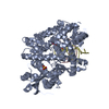
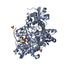
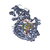
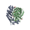
| Title | Crystal structure of the monomeric isocitrate dehydrogenase in complex with isocitrate and Mn | ||||||
 Components Components | Isocitrate dehydrogenase | ||||||
 Keywords Keywords | OXIDOREDUCTASE / Greece key motif | ||||||
| Function / homology |  Function and homology information Function and homology informationisocitrate dehydrogenase (NADP+) / isocitrate dehydrogenase (NADP+) activity / glyoxylate cycle / tricarboxylic acid cycle / metal ion binding / cytoplasm Similarity search - Function | ||||||
| Biological species |  Azotobacter vinelandii (bacteria) Azotobacter vinelandii (bacteria) | ||||||
| Method |  X-RAY DIFFRACTION / SYNCHROTRON / MAD, Molecular Replacement method / Resolution: 1.95 Å X-RAY DIFFRACTION / SYNCHROTRON / MAD, Molecular Replacement method / Resolution: 1.95 Å | ||||||
 Authors Authors | Yasutake, Y. / Watanabe, S. / Yao, M. / Takada, Y. / Fukunaga, N. / Tanaka, I. | ||||||
 Citation Citation | Journal: Structure / Year: 2002 Title: Structure of the Monomeric Isocitrate Dehydrogenase: Evidence of a Protein Monomerization by a Domain Duplication Authors: Yasutake, Y. / Watanabe, S. / Yao, M. / Takada, Y. / Fukunaga, N. / Tanaka, I. #1: Journal: Acta Crystallogr.,Sect.D / Year: 2001Title: Crystallization and preliminary X-ray diffraction studies of monomeric isocitrate dehydrogenase by the MAD method using Mn atoms Authors: Yasutake, Y. / Watanabe, S. / Yao, M. / Takada, Y. / Fukunaga, N. / Tanaka, I. | ||||||
| History |
|
- Structure visualization
| Structure viewer | Molecule: MolmilJmol/JSmol |
|---|
- Downloads & links
-Download
| PDBx/mmCIF format | 1itw.cif.gz | 603.6 KB | Display | PDBx/mmCIF format |
|---|---|---|---|---|
| PDB format | pdb1itw.ent.gz | 491.9 KB | Display | PDB format |
| PDBx/mmJSON format | 1itw.json.gz | Tree view | PDBx/mmJSON format | |
| Others |  Other downloads Other downloads |
-Validation report
| Arichive directory | https://data.pdbj.org/pub/pdb/validation_reports/it/1itwftp://data.pdbj.org/pub/pdb/validation_reports/it/1itw | HTTPS FTP |
|---|
-Related structure data
| Similar structure data |
|---|


-Links
 PDBj
PDBj


- Assembly
| Deposited unit | 
| ||||||||
|---|---|---|---|---|---|---|---|---|---|
| 1 | 
| ||||||||
| 2 | 
| ||||||||
| 3 | 
| ||||||||
| 4 | 
| ||||||||
| Unit cell |
|
-Components
| #1: Protein | Mass: 80507.633 Da / Num. of mol.: 4 / Source method: isolated from a natural source / Source: (natural) Azotobacter vinelandii (bacteria) / Strain: IAM1078References: UniProt: P16100, isocitrate dehydrogenase (NADP+) #2: Chemical | ChemComp-MN /   Mass: 54.938 Da / Num. of mol.: 5 / Source method: obtained synthetically / Formula: Mn Mass: 54.938 Da / Num. of mol.: 5 / Source method: obtained synthetically / Formula: Mn#3: Chemical | ChemComp-ICT /   Mass: 192.124 Da / Num. of mol.: 4 / Source method: obtained synthetically / Formula: C6H8O7 Mass: 192.124 Da / Num. of mol.: 4 / Source method: obtained synthetically / Formula: C6H8O7#4: Water | ChemComp-HOH / |  Mass: 18.015 Da / Num. of mol.: 2306 / Source method: isolated from a natural source / Formula: H2O Mass: 18.015 Da / Num. of mol.: 2306 / Source method: isolated from a natural source / Formula: H2O |
|---|
-Experimental details
-Experiment
| Experiment | Method: X-RAY DIFFRACTION / Number of used crystals: 1 |
|---|
- Sample preparation
| Crystal | Density Matthews: 2.6 Å3/Da / Density % sol: 52.2 % | |||||||||||||||||||||||||||||||||||||||||||||||||||||||||||||||
|---|---|---|---|---|---|---|---|---|---|---|---|---|---|---|---|---|---|---|---|---|---|---|---|---|---|---|---|---|---|---|---|---|---|---|---|---|---|---|---|---|---|---|---|---|---|---|---|---|---|---|---|---|---|---|---|---|---|---|---|---|---|---|---|---|
| Crystal grow | Temperature: 293 K / Method: vapor diffusion, hanging drop / pH: 7 Details: HEPES, PEG6000, glycerol, manganese chloride, DL-isocitrate, pH 7.0, VAPOR DIFFUSION, HANGING DROP, temperature 293K | |||||||||||||||||||||||||||||||||||||||||||||||||||||||||||||||
| Crystal grow | *PLUS Temperature: 291 KDetails: Yasutake, Y., (2001) Acta Crystallogr., Sect.D, 57, 1682. | |||||||||||||||||||||||||||||||||||||||||||||||||||||||||||||||
| Components of the solutions | *PLUS
|
-Data collection
| Diffraction | Mean temperature: 100 K |
|---|---|
| Diffraction source | Source: SYNCHROTRON / Site: SPring-8  / Beamline: BL41XU / Wavelength: 0.9 Å / Beamline: BL41XU / Wavelength: 0.9 Å |
| Detector | Type: MARRESEARCH / Detector: CCD / Date: Sep 20, 2001 |
| Radiation | Monochromator: MIRROR / Protocol: SINGLE WAVELENGTH / Monochromatic (M) / Laue (L): M / Scattering type: x-ray |
| Radiation wavelength | Wavelength: 0.9 Å / Relative weight: 1 |
| Reflection | Resolution: 1.95→20 Å / Num. all: 237767 / Num. obs: 235314 / % possible obs: 98.9 % / Observed criterion σ(I): 3 / Redundancy: 3.6 % / Biso Wilson estimate: 24.474 Å2 / Rmerge(I) obs: 0.083 / Rsym value: 0.071 / Net I/σ(I): 8.6 |
| Reflection shell | Resolution: 1.95→2.06 Å / Redundancy: 3.2 % / Rmerge(I) obs: 0.388 / Mean I/σ(I) obs: 2 / Num. unique all: 33505 / Rsym value: 0.327 / % possible all: 96.7 |
| Reflection | *PLUS Lowest resolution: 40 Å / Num. measured all: 846014 |
| Reflection shell | *PLUS % possible obs: 96.7 % |
- Processing
| Software |
| ||||||||||||||||||||||||||||||||||||
|---|---|---|---|---|---|---|---|---|---|---|---|---|---|---|---|---|---|---|---|---|---|---|---|---|---|---|---|---|---|---|---|---|---|---|---|---|---|
| Refinement | Method to determine structure: MAD, Molecular Replacement method Resolution: 1.95→10 Å / Isotropic thermal model: Isotropic for overall / Cross valid method: THROUGHOUT / σ(F): 0 / Stereochemistry target values: Engh & Huber
| ||||||||||||||||||||||||||||||||||||
| Solvent computation | Solvent model: throughout / Bsol: 69 Å2 / ksol: 0.5 e/Å3 | ||||||||||||||||||||||||||||||||||||
| Displacement parameters | Biso mean: 23.5096 Å2
| ||||||||||||||||||||||||||||||||||||
| Refine analyze |
| ||||||||||||||||||||||||||||||||||||
| Refinement step | Cycle: LAST / Resolution: 1.95→10 Å
| ||||||||||||||||||||||||||||||||||||
| Refine LS restraints |
| ||||||||||||||||||||||||||||||||||||
| LS refinement shell | Resolution: 1.95→2.02 Å / Total num. of bins used: 10
| ||||||||||||||||||||||||||||||||||||
| Xplor file | Serial no: 1 / Param file: protein_rep.param / Topol file: protein.top | ||||||||||||||||||||||||||||||||||||
| Refinement | *PLUS Lowest resolution: 10 Å / Num. reflection obs: 233612 / % reflection Rfree: 10 % | ||||||||||||||||||||||||||||||||||||
| Solvent computation | *PLUS | ||||||||||||||||||||||||||||||||||||
| Displacement parameters | *PLUS | ||||||||||||||||||||||||||||||||||||
| Refine LS restraints | *PLUS
|
