 Movie
Movie Controller

[English] 日本語
 Yorodumi
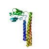
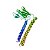
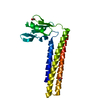
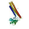
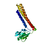
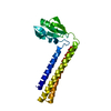
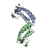
- PDB-1is1: Crystal structure of ribosome recycling factor from Vibrio paraha... -
+ Open data

- Basic information
| Entry | Database: PDB / ID: 1is1 | ||||||
|---|---|---|---|---|---|---|---|
| Title | Crystal structure of ribosome recycling factor from Vibrio parahaemolyticus | ||||||
 Components Components | RIBOSOME RECYCLING FACTOR | ||||||
 Keywords Keywords | TRANSLATION | ||||||
| Function / homology |  Function and homology information Function and homology informationcytoplasmic translational termination / ribosomal large subunit binding / cytosol Similarity search - Function | ||||||
| Biological species |   Vibrio parahaemolyticus (bacteria) Vibrio parahaemolyticus (bacteria) | ||||||
| Method |  X-RAY DIFFRACTION / SYNCHROTRON / MOLECULAR REPLACEMENT / Resolution: 2.2 Å X-RAY DIFFRACTION / SYNCHROTRON / MOLECULAR REPLACEMENT / Resolution: 2.2 Å | ||||||
 Authors Authors | Nakano, H. / Yamaichi, Y. / Uchiyama, S. / Yoshida, T. / Nishina, K. / Kato, H. / Ohkubo, T. / Honda, T. / Yamagata, Y. / Kobayashi, Y. | ||||||
 Citation Citation | Journal: J.BIOL.CHEM. / Year: 2003 Title: Structure and binding mode of a ribosome recycling factor (RRF) from mesophilic bacterium Authors: Nakano, H. / Yoshida, T. / Uchiyama, S. / Kawachi, M. / Matsuo, H. / Kato, T. / Ohshima, A. / Yamaichi, Y. / Honda, T. / Kato, H. / Yamagata, Y. / Ohkubo, T. / Kobayashi, Y. | ||||||
| History |
|
- Structure visualization
| Structure viewer | Molecule: MolmilJmol/JSmol |
|---|
- Downloads & links
-Download
| PDBx/mmCIF format | 1is1.cif.gz | 51.3 KB | Display | PDBx/mmCIF format |
|---|---|---|---|---|
| PDB format | pdb1is1.ent.gz | 37.7 KB | Display | PDB format |
| PDBx/mmJSON format | 1is1.json.gz | Tree view | PDBx/mmJSON format | |
| Others |  Other downloads Other downloads |
-Validation report
| Arichive directory | https://data.pdbj.org/pub/pdb/validation_reports/is/1is1ftp://data.pdbj.org/pub/pdb/validation_reports/is/1is1 | HTTPS FTP |
|---|
-Related structure data
| Similar structure data |
|---|


-Links
 PDBj
PDBj- Assembly
| Deposited unit | 
| |||||||||||||||
|---|---|---|---|---|---|---|---|---|---|---|---|---|---|---|---|---|
| 1 |
| |||||||||||||||
| Unit cell |
| |||||||||||||||
| Components on special symmetry positions |
|
-Components
| #1: Protein | Mass: 20635.791 Da / Num. of mol.: 1 Source method: isolated from a genetically manipulated source Source: (gene. exp.) Vibrio parahaemolyticus (bacteria) / Plasmid: pET22B / Production host: |
|---|---|
| #2: Water | ChemComp-HOH /  Mass: 18.015 Da / Num. of mol.: 227 / Source method: isolated from a natural source / Formula: H2O Mass: 18.015 Da / Num. of mol.: 227 / Source method: isolated from a natural source / Formula: H2O |
-Experimental details
-Experiment
| Experiment | Method: X-RAY DIFFRACTION / Number of used crystals: 1 |
|---|
- Sample preparation
| Crystal | Density Matthews: 2.39 Å3/Da / Density % sol: 48.58 % | ||||||||||||||||||||||||||||||
|---|---|---|---|---|---|---|---|---|---|---|---|---|---|---|---|---|---|---|---|---|---|---|---|---|---|---|---|---|---|---|---|
| Crystal grow | Temperature: 277 K / Method: vapor diffusion, hanging drop / pH: 6.2 Details: PEG 8000, sodium acetate, GDP, fusidic acid, pH 6.2, VAPOR DIFFUSION, HANGING DROP, temperature 277K | ||||||||||||||||||||||||||||||
| Crystal grow | *PLUS Temperature: 4 ℃ / Method: vapor diffusion, hanging drop | ||||||||||||||||||||||||||||||
| Components of the solutions | *PLUS
|
-Data collection
| Diffraction | Mean temperature: 100 K |
|---|---|
| Diffraction source | Source: SYNCHROTRON / Site: Photon Factory  / Beamline: BL-6A / Wavelength: 1 Å / Beamline: BL-6A / Wavelength: 1 Å |
| Detector | Type: WEISSENBERG / Detector: DIFFRACTOMETER |
| Radiation | Protocol: SINGLE WAVELENGTH / Monochromatic (M) / Laue (L): M / Scattering type: x-ray |
| Radiation wavelength | Wavelength: 1 Å / Relative weight: 1 |
| Reflection | Resolution: 2.2→40 Å / Num. obs: 10326 / % possible obs: 99.6 % / Observed criterion σ(F): 2 / Observed criterion σ(I): 2 / Rmerge(I) obs: 0.073 |
| Reflection shell | Resolution: 2.2→2.24 Å / % possible all: 93.7 |
| Reflection | *PLUS Highest resolution: 2.2 Å / Num. measured all: 82069 |
| Reflection shell | *PLUS Rmerge(I) obs: 0.251 |
- Processing
| Software |
| |||||||||||||||||||||||||
|---|---|---|---|---|---|---|---|---|---|---|---|---|---|---|---|---|---|---|---|---|---|---|---|---|---|---|
| Refinement | Method to determine structure: MOLECULAR REPLACEMENT / Resolution: 2.2→20 Å / σ(F): 2 / Stereochemistry target values: Engh & Huber
| |||||||||||||||||||||||||
| Refinement step | Cycle: LAST / Resolution: 2.2→20 Å
| |||||||||||||||||||||||||
| Refinement | *PLUS Highest resolution: 2.2 Å / Num. reflection obs: 9947 / % reflection Rfree: 5 % | |||||||||||||||||||||||||
| Solvent computation | *PLUS | |||||||||||||||||||||||||
| Displacement parameters | *PLUS | |||||||||||||||||||||||||
| Refine LS restraints | *PLUS
| |||||||||||||||||||||||||
| LS refinement shell | *PLUS Highest resolution: 2.2 Å / Lowest resolution: 2.3 Å / Rfactor Rfree: 0.278 / Rfactor Rwork: 0.207 |
