 Movie
Movie Controller

[English] 日本語
 Yorodumi
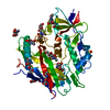
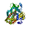
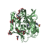
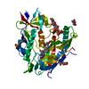
- PDB-1io8: Thermophilic cytochrome P450 (CYP119) from sulfolobus solfataricu... -
+ Open data

- Basic information
| Entry | Database: PDB / ID: 1io8 | ||||||
|---|---|---|---|---|---|---|---|
| Title | Thermophilic cytochrome P450 (CYP119) from sulfolobus solfataricus: High resolution structural origin of its thermostability and functional properties | ||||||
 Components Components | CYTOCHROME P450 CYP119 | ||||||
 Keywords Keywords | OXIDOREDUCTASE / Thermophilic / Cytochromo P450 / RIKEN Structural Genomics/Proteomics Initiative / RSGI / Structural Genomics / NPPSFA / National Project on Protein Structural and Functional Analyses | ||||||
| Function / homology |  Function and homology information Function and homology informationOxidoreductases; Acting on paired donors, with incorporation or reduction of molecular oxygen / lactoperoxidase activity / peroxidase / oxidoreductase activity, acting on paired donors, with incorporation or reduction of molecular oxygen / monooxygenase activity / iron ion binding / heme binding / cytoplasm Similarity search - Function | ||||||
| Biological species |   Sulfolobus solfataricus (archaea) Sulfolobus solfataricus (archaea) | ||||||
| Method |  X-RAY DIFFRACTION / SYNCHROTRON / MOLECULAR REPLACEMENT / Resolution: 2 Å X-RAY DIFFRACTION / SYNCHROTRON / MOLECULAR REPLACEMENT / Resolution: 2 Å | ||||||
 Authors Authors | Park, S.-Y. / Yamane, K. / Adachi, S. / Shiro, Y. / Sligar, S.G. / RIKEN Structural Genomics/Proteomics Initiative (RSGI) | ||||||
 Citation Citation | Journal: J.Inorg.Biochem. / Year: 2002 Title: Thermophilic cytochrome P450 (CYP119) from Sulfolobus solfataricus: high resolution structure and functional properties Authors: Park, S.Y. / Yamane, K. / Adachi, S. / Shiro, Y. / Weiss, K.E. / Maves, S.A. / Sligar, S.G. | ||||||
| History |
|
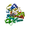
- Structure visualization
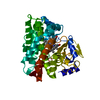
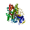
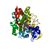
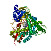
| Structure viewer | Molecule: MolmilJmol/JSmol |
|---|
- Downloads & links
-Download
| PDBx/mmCIF format | 1io8.cif.gz | 166.7 KB | Display | PDBx/mmCIF format |
|---|---|---|---|---|
| PDB format | pdb1io8.ent.gz | 132.2 KB | Display | PDB format |
| PDBx/mmJSON format | 1io8.json.gz | Tree view | PDBx/mmJSON format | |
| Others |  Other downloads Other downloads |
-Validation report
| Arichive directory | https://data.pdbj.org/pub/pdb/validation_reports/io/1io8ftp://data.pdbj.org/pub/pdb/validation_reports/io/1io8 | HTTPS FTP |
|---|
-Related structure data


-Links
 PDBj
PDBj
- Assembly
| Deposited unit | 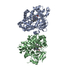
| ||||||||
|---|---|---|---|---|---|---|---|---|---|
| 1 | 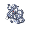
| ||||||||
| 2 | 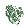
| ||||||||
| 3 | 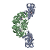
| ||||||||
| Unit cell |
|
-Components
| #1: Protein | Mass: 42890.012 Da / Num. of mol.: 2 / Mutation: F24L Source method: isolated from a genetically manipulated source Source: (gene. exp.) Sulfolobus solfataricus (archaea) / Plasmid: PKWS119 / Production host:  References: UniProt: Q55080, Oxidoreductases; Acting on paired donors, with incorporation or reduction of molecular oxygen; With reduced flavin or flavoprotein as one donor, and incorporation of one ...References: UniProt: Q55080, Oxidoreductases; Acting on paired donors, with incorporation or reduction of molecular oxygen; With reduced flavin or flavoprotein as one donor, and incorporation of one atom of oxygen into the other donor #2: Chemical |   Mass: 616.487 Da / Num. of mol.: 2 / Source method: obtained synthetically / Formula: C34H32FeN4O4 Mass: 616.487 Da / Num. of mol.: 2 / Source method: obtained synthetically / Formula: C34H32FeN4O4#3: Water | ChemComp-HOH / |  Mass: 18.015 Da / Num. of mol.: 283 / Source method: isolated from a natural source / Formula: H2O Mass: 18.015 Da / Num. of mol.: 283 / Source method: isolated from a natural source / Formula: H2O |
|---|
-Experimental details
-Experiment
| Experiment | Method: X-RAY DIFFRACTION / Number of used crystals: 1 |
|---|
- Sample preparation
| Crystal | Density Matthews: 2.36 Å3/Da / Density % sol: 47.87 % | ||||||||||||||||||||
|---|---|---|---|---|---|---|---|---|---|---|---|---|---|---|---|---|---|---|---|---|---|
| Crystal grow | Temperature: 298 K / Method: vapor diffusion, sitting drop / pH: 5.6 Details: PEG4000, 0.2M sodium thiocyanate, pH 5.6, VAPOR DIFFUSION, SITTING DROP, temperature 298K | ||||||||||||||||||||
| Crystal | *PLUS Density % sol: 48 % | ||||||||||||||||||||
| Crystal grow | *PLUS Temperature: 293 K / pH: 6.4 / Details: Park, S.Y., (2000) Acta Crystallogr., D56, 1173. | ||||||||||||||||||||
| Components of the solutions | *PLUS
|
-Data collection
| Diffraction | Mean temperature: 298 K |
|---|---|
| Diffraction source | Source: SYNCHROTRON / Site: SPring-8  / Beamline: BL44B2 / Wavelength: 0.7 Å / Beamline: BL44B2 / Wavelength: 0.7 Å |
| Detector | Type: MARRESEARCH / Detector: CCD / Details: mirrors |
| Radiation | Monochromator: Si 111 / Protocol: SINGLE WAVELENGTH / Monochromatic (M) / Laue (L): M / Scattering type: x-ray |
| Radiation wavelength | Wavelength: 0.7 Å / Relative weight: 1 |
| Reflection | Resolution: 2→20 Å / Num. all: 188348 / Num. obs: 53362 / % possible obs: 94.9 % / Observed criterion σ(I): 0 / Redundancy: 3.5 % / Biso Wilson estimate: 21.6 Å2 / Rmerge(I) obs: 0.056 / Net I/σ(I): 16.7 |
| Reflection shell | Resolution: 2→2.07 Å / Redundancy: 3.2 % / Rmerge(I) obs: 0.463 / Mean I/σ(I) obs: 2.6 / % possible all: 86.2 |
| Reflection | *PLUS Num. measured all: 188348 |
| Reflection shell | *PLUS % possible obs: 86.2 % |
- Processing
| Software |
| |||||||||||||||||||||||||
|---|---|---|---|---|---|---|---|---|---|---|---|---|---|---|---|---|---|---|---|---|---|---|---|---|---|---|
| Refinement | Method to determine structure: MOLECULAR REPLACEMENT Starting model: Native CYP119 Resolution: 2→20 Å / Isotropic thermal model: Isotropic / Cross valid method: THROUGHOUT / σ(F): 0 / Stereochemistry target values: Engh & Huber
| |||||||||||||||||||||||||
| Displacement parameters | Biso mean: 35 Å2 | |||||||||||||||||||||||||
| Refine analyze |
| |||||||||||||||||||||||||
| Refinement step | Cycle: LAST / Resolution: 2→20 Å
| |||||||||||||||||||||||||
| Refine LS restraints |
| |||||||||||||||||||||||||
| LS refinement shell | Resolution: 2→2.13 Å
| |||||||||||||||||||||||||
| Software | *PLUS Name: X-PLOR / Version: 3.851 / Classification: refinement | |||||||||||||||||||||||||
| Refinement | *PLUS Highest resolution: 2 Å / Lowest resolution: 20 Å / σ(F): 0 / % reflection Rfree: 5 % / Rfactor obs: 0.228 / Rfactor Rwork: 0.23 | |||||||||||||||||||||||||
| Solvent computation | *PLUS | |||||||||||||||||||||||||
| Displacement parameters | *PLUS Biso mean: 35 Å2 | |||||||||||||||||||||||||
| Refine LS restraints | *PLUS
| |||||||||||||||||||||||||
| LS refinement shell | *PLUS Highest resolution: 2 Å / Rfactor Rfree: 0.373 / Rfactor Rwork: 0.388 |
