 Movie
Movie Controller

[English] 日本語
 Yorodumi
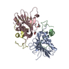
- PDB-1imd: STRUCTURAL STUDIES OF METAL BINDING BY INOSITOL MONOPHOSPHATASE: ... -
+ Open data

- Basic information
| Entry | Database: PDB / ID: 1imd | ||||||
|---|---|---|---|---|---|---|---|
| Title | STRUCTURAL STUDIES OF METAL BINDING BY INOSITOL MONOPHOSPHATASE: EVIDENCE FOR TWO-METAL ION CATALYSIS | ||||||
 Components Components | INOSITOL MONOPHOSPHATASE | ||||||
 Keywords Keywords | HYDROLASE | ||||||
| Function / homology |  Function and homology information Function and homology informationD-galactose 1-phosphate phosphatase / inositol monophosphate phosphatase activity / glucose-1-phosphatase activity / glycerol-2-phosphatase activity / glucose-6-phosphatase activity / inositol monophosphate 4-phosphatase activity / fructose-1-phosphatase activity / inositol monophosphate 3-phosphatase activity / Synthesis of IP2, IP, and Ins in the cytosol / lithium ion binding ...D-galactose 1-phosphate phosphatase / inositol monophosphate phosphatase activity / glucose-1-phosphatase activity / glycerol-2-phosphatase activity / glucose-6-phosphatase activity / inositol monophosphate 4-phosphatase activity / fructose-1-phosphatase activity / inositol monophosphate 3-phosphatase activity / Synthesis of IP2, IP, and Ins in the cytosol / lithium ion binding / inositol biosynthetic process / inositol-phosphate phosphatase / inositol monophosphate 1-phosphatase activity / inositol metabolic process / phosphatidylinositol biosynthetic process / phosphate-containing compound metabolic process / phosphatidylinositol phosphate biosynthetic process / manganese ion binding / magnesium ion binding / signal transduction / protein homodimerization activity / identical protein binding / cytosol / cytoplasm Similarity search - Function | ||||||
| Biological species |  Homo sapiens (human) Homo sapiens (human) | ||||||
| Method |  X-RAY DIFFRACTION / Resolution: 2.6 Å X-RAY DIFFRACTION / Resolution: 2.6 Å | ||||||
 Authors Authors | Bone, R. | ||||||
 Citation Citation | Journal: Biochemistry / Year: 1994 Title: Structural studies of metal binding by inositol monophosphatase: evidence for two-metal ion catalysis. Authors: Bone, R. / Frank, L. / Springer, J.P. / Atack, J.R. #1: Journal: Biochem.J. / Year: 1992Title: Cdna Cloning of Human and Rat Brain Myo-Inositol Monophosphatase. Expression and Characterization of the Human Recombinant Enzyme Authors: Mcallister, G. / Whiting, P. / Hammond, E.A. / Knowles, M.R. / Atack, J.R. / Bailey, F.J. / Maigetter, R. / Ragan, C.I. #2: Journal: Proc.Natl.Acad.Sci.USA / Year: 1992Title: Structure of Inositol Monophosphatase, the Putative Target of Lithium Therapy Authors: Bone, R. / Springer, J.P. / Atack, J.R. | ||||||
| History |
|
- Structure visualization
| Structure viewer | Molecule: MolmilJmol/JSmol |
|---|
- Downloads & links
-Download
| PDBx/mmCIF format | 1imd.cif.gz | 114.1 KB | Display | PDBx/mmCIF format |
|---|---|---|---|---|
| PDB format | pdb1imd.ent.gz | 89.4 KB | Display | PDB format |
| PDBx/mmJSON format | 1imd.json.gz | Tree view | PDBx/mmJSON format | |
| Others |  Other downloads Other downloads |
-Validation report
| Arichive directory | https://data.pdbj.org/pub/pdb/validation_reports/im/1imdftp://data.pdbj.org/pub/pdb/validation_reports/im/1imd | HTTPS FTP |
|---|
-Related structure data


-Links
 PDBj
PDBj- Assembly
| Deposited unit | 
| ||||||||
|---|---|---|---|---|---|---|---|---|---|
| 1 |
| ||||||||
| Unit cell |
| ||||||||
| Atom site foot note | 1: TYR A 62 - PRO A 63 OMEGA = 211.14 PEPTIDE BOND DEVIATES SIGNIFICANTLY FROM TRANS CONFORMATION 2: CIS PROLINE - PRO A 186 3: PHE B 102 - PRO B 103 OMEGA = 211.75 PEPTIDE BOND DEVIATES SIGNIFICANTLY FROM TRANS CONFORMATION 4: CIS PROLINE - PRO B 186 |
-Components
| #1: Protein | Mass: 30219.781 Da / Num. of mol.: 2 Source method: isolated from a genetically manipulated source Source: (gene. exp.) Homo sapiens (human) / Gene: CDNA / Organ: BRAIN / References: UniProt: P29218, inositol-phosphate phosphatase#2: Chemical | ChemComp-MN /   Mass: 54.938 Da / Num. of mol.: 4 / Source method: obtained synthetically / Formula: Mn Mass: 54.938 Da / Num. of mol.: 4 / Source method: obtained synthetically / Formula: Mn#3: Chemical |   Mass: 94.971 Da / Num. of mol.: 2 / Source method: obtained synthetically / Formula: PO4 Mass: 94.971 Da / Num. of mol.: 2 / Source method: obtained synthetically / Formula: PO4#4: Water | ChemComp-HOH / |  Mass: 18.015 Da / Num. of mol.: 50 / Source method: isolated from a natural source / Formula: H2O Mass: 18.015 Da / Num. of mol.: 50 / Source method: isolated from a natural source / Formula: H2OHas protein modification | Y | |
|---|
-Experimental details
-Experiment
| Experiment | Method: X-RAY DIFFRACTION |
|---|
- Sample preparation
| Crystal | Density Matthews: 2.7 Å3/Da / Density % sol: 54.49 % | ||||||||||||||||||||||||||||||||||||||||||||||||||||||||||||
|---|---|---|---|---|---|---|---|---|---|---|---|---|---|---|---|---|---|---|---|---|---|---|---|---|---|---|---|---|---|---|---|---|---|---|---|---|---|---|---|---|---|---|---|---|---|---|---|---|---|---|---|---|---|---|---|---|---|---|---|---|---|
| Crystal grow | *PLUS pH: 7.5 / Method: vapor diffusion | ||||||||||||||||||||||||||||||||||||||||||||||||||||||||||||
| Components of the solutions | *PLUS
|
-Data collection
| Radiation | Scattering type: x-ray |
|---|---|
| Radiation wavelength | Relative weight: 1 |
| Reflection | *PLUS Highest resolution: 2.6 Å / Lowest resolution: 8 Å / % possible obs: 95 % / Rmerge(I) obs: 0.083 |
- Processing
| Software |
| ||||||||||||||||||||||||||||||||||||||||||||||||||||||||||||
|---|---|---|---|---|---|---|---|---|---|---|---|---|---|---|---|---|---|---|---|---|---|---|---|---|---|---|---|---|---|---|---|---|---|---|---|---|---|---|---|---|---|---|---|---|---|---|---|---|---|---|---|---|---|---|---|---|---|---|---|---|---|
| Refinement | Resolution: 2.6→8 Å / Rfactor Rwork: 0.188 / Rfactor obs: 0.188 / σ(F): 0 | ||||||||||||||||||||||||||||||||||||||||||||||||||||||||||||
| Refinement step | Cycle: LAST / Resolution: 2.6→8 Å
| ||||||||||||||||||||||||||||||||||||||||||||||||||||||||||||
| Refine LS restraints |
|
