 Movie
Movie Controller

[English] 日本語
 Yorodumi
- PDB-1ilt: X-RAY STRUCTURE OF INTERLEUKIN-1 RECEPTOR ANTAGONIST AT 2.0 ANGST... -
+ Open data

- Basic information
| Entry | Database: PDB / ID: 1ilt | ||||||
|---|---|---|---|---|---|---|---|
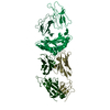
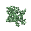
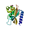
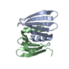
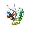
| Title | X-RAY STRUCTURE OF INTERLEUKIN-1 RECEPTOR ANTAGONIST AT 2.0 ANGSTROMS RESOLUTION | ||||||
 Components Components | INTERLEUKIN-1 RECEPTOR ANTAGONIST | ||||||
 Keywords Keywords | CYTOKINE | ||||||
| Function / homology |  Function and homology information Function and homology informationinterleukin-1 type I receptor antagonist activity / interleukin-1 type II receptor antagonist activity / interleukin-1, type I receptor binding / interleukin-1 receptor antagonist activity / interleukin-1, type II receptor binding / negative regulation of interleukin-1-mediated signaling pathway / negative regulation of heterotypic cell-cell adhesion / interleukin-1 receptor binding / insulin secretion / Interleukin-10 signaling ...interleukin-1 type I receptor antagonist activity / interleukin-1 type II receptor antagonist activity / interleukin-1, type I receptor binding / interleukin-1 receptor antagonist activity / interleukin-1, type II receptor binding / negative regulation of interleukin-1-mediated signaling pathway / negative regulation of heterotypic cell-cell adhesion / interleukin-1 receptor binding / insulin secretion / Interleukin-10 signaling / response to glucocorticoid / cytokine activity / acute-phase response / lipid metabolic process / Interleukin-1 signaling / immune response / inflammatory response / centrosome / : / extracellular exosome / nucleoplasm / plasma membrane / cytosol Similarity search - Function | ||||||
| Biological species |  Homo sapiens (human) Homo sapiens (human) | ||||||
| Method |  X-RAY DIFFRACTION / Resolution: 2 Å X-RAY DIFFRACTION / Resolution: 2 Å | ||||||
 Authors Authors | Brandhuber, B.J. / Vigers, G.P.A. | ||||||
 Citation Citation | Journal: J.Biol.Chem. / Year: 1994 Title: X-ray structure of interleukin-1 receptor antagonist at 2.0-A resolution. Authors: Vigers, G.P. / Caffes, P. / Evans, R.J. / Thompson, R.C. / Eisenberg, S.P. / Brandhuber, B.J. | ||||||
| History |
| ||||||
| Remark 700 | SHEET THE SHEETS PRESENTED AS A1 AND B1 ON SHEET RECORDS BELOW ARE ACTUALLY A SIX-STRANDED BETA- ...SHEET THE SHEETS PRESENTED AS A1 AND B1 ON SHEET RECORDS BELOW ARE ACTUALLY A SIX-STRANDED BETA-BARREL. THIS IS REPRESENTED BY A SEVEN-STRANDED SHEET IN WHICH THE FIRST AND LAST STRANDS ARE IDENTICAL. |
- Structure visualization
| Structure viewer | Molecule: MolmilJmol/JSmol |
|---|
- Downloads & links
-Download
| PDBx/mmCIF format | 1ilt.cif.gz | 20.4 KB | Display | PDBx/mmCIF format |
|---|---|---|---|---|
| PDB format | pdb1ilt.ent.gz | 10.5 KB | Display | PDB format |
| PDBx/mmJSON format | 1ilt.json.gz | Tree view | PDBx/mmJSON format | |
| Others |  Other downloads Other downloads |
-Validation report
| Arichive directory | https://data.pdbj.org/pub/pdb/validation_reports/il/1iltftp://data.pdbj.org/pub/pdb/validation_reports/il/1ilt | HTTPS FTP |
|---|
-Related structure data
| Similar structure data |
|---|


-Links
 PDBj
PDBj


- Assembly
| Deposited unit | 
| ||||||||
|---|---|---|---|---|---|---|---|---|---|
| 1 | 
| ||||||||
| 2 | 
| ||||||||
| Unit cell |
| ||||||||
| Atom site foot note | 1: CIS PROLINE - PRO A 53 / 2: CIS PROLINE - PRO B 53 | ||||||||
| Noncrystallographic symmetry (NCS) | NCS oper: (Code: given Matrix: (-0.530352, 0.829762, 0.173845), Vector: Details | TWO INDEPENDENT MOLECULES PER UNIT CELL. THE TRANSFORMATION PRESENTED ON *MTRIX* RECORDS BELOW WILL YIELD APPROXIMATE COORDINATES FOR CHAIN *B* WHEN APPLIED TO CHAIN *A*. | |
-Components
| #1: Protein | Mass: 17145.406 Da / Num. of mol.: 2 Source method: isolated from a genetically manipulated source Source: (gene. exp.) Homo sapiens (human) / References: UniProt: P18510 |
|---|
-Experimental details
-Experiment
| Experiment | Method: X-RAY DIFFRACTION |
|---|
- Sample preparation
| Crystal | Density Matthews: 2.07 Å3/Da / Density % sol: 40.72 % | ||||||||||||||||||||||||||||||||||||||||||
|---|---|---|---|---|---|---|---|---|---|---|---|---|---|---|---|---|---|---|---|---|---|---|---|---|---|---|---|---|---|---|---|---|---|---|---|---|---|---|---|---|---|---|---|
| Crystal grow | *PLUS Temperature: 20 ℃ / pH: 6 / Method: vapor diffusion, hanging dropDetails: drop contained 1:1 mixture of precipitant and recombinant IL-lra | ||||||||||||||||||||||||||||||||||||||||||
| Components of the solutions | *PLUS
|
-Data collection
| Radiation | Scattering type: x-ray |
|---|---|
| Radiation wavelength | Relative weight: 1 |
| Reflection | *PLUS Highest resolution: 2 Å / Lowest resolution: 8 Å / % possible obs: 95.6 % / Rmerge(I) obs: 0.069 |
- Processing
| Software |
| ||||||||||||||||||||||||||||||||||||||||||||||||||||||||||||
|---|---|---|---|---|---|---|---|---|---|---|---|---|---|---|---|---|---|---|---|---|---|---|---|---|---|---|---|---|---|---|---|---|---|---|---|---|---|---|---|---|---|---|---|---|---|---|---|---|---|---|---|---|---|---|---|---|---|---|---|---|---|
| Refinement | Resolution: 2→8 Å / σ(F): 2 /
| ||||||||||||||||||||||||||||||||||||||||||||||||||||||||||||
| Refinement step | Cycle: LAST / Resolution: 2→8 Å
| ||||||||||||||||||||||||||||||||||||||||||||||||||||||||||||
| Refine LS restraints |
| ||||||||||||||||||||||||||||||||||||||||||||||||||||||||||||
| Refinement | *PLUS | ||||||||||||||||||||||||||||||||||||||||||||||||||||||||||||
| Solvent computation | *PLUS | ||||||||||||||||||||||||||||||||||||||||||||||||||||||||||||
| Displacement parameters | *PLUS | ||||||||||||||||||||||||||||||||||||||||||||||||||||||||||||
| Refine LS restraints | *PLUS Type: x_angle_deg / Dev ideal: 1.9 |
