 Movie
Movie Controller

[English] 日本語
 Yorodumi
- PDB-1ijc: Solution Structure of Bucandin, a Neurotoxin from the Venom of th... -
+ Open data

- Basic information
| Entry | Database: PDB / ID: 1ijc | ||||||
|---|---|---|---|---|---|---|---|
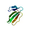
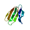
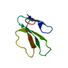
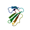
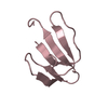
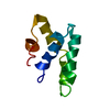
| Title | Solution Structure of Bucandin, a Neurotoxin from the Venom of the Malayan Krait | ||||||
 Components Components | bucandin | ||||||
 Keywords Keywords | TOXIN / three-finger motif / two antiparallel beta-sheets / two and four stranded beta-sheets | ||||||
| Function / homology |  Function and homology information Function and homology information | ||||||
| Biological species |  Bungarus candidus (cobra) Bungarus candidus (cobra) | ||||||
| Method | SOLUTION NMR / distance geometry, simulated annealing, molecular dynamics, torsion angle dynamics | ||||||
 Authors Authors | Torres, A.M. / Kini, R.M. / Nirthanan, S. / Kuchel, P.W. | ||||||
 Citation Citation | Journal: Biochem.J. / Year: 2001 Title: NMR structure of bucandin, a neurotoxin from the venom of the Malayan krait (Bungarus candidus). Authors: Torres, A.M. / Kini, R.M. / Selvanayagam, N. / Kuchel, P.W. #1: Journal: Acta Crystallogr.,Sect.D / Year: 2000Title: The atomic resolution structure of bucandin, a novel toxin isolated from the Malayan krait, determined by direct methods Authors: Kuhn, P. / Deacon, A.M. / Comoso, S. / Rajaseger, G. / Kini, R.M. / Uson, I. / Kolatkar, P.R. | ||||||
| History |
|
- Structure visualization
| Structure viewer | Molecule: MolmilJmol/JSmol |
|---|
- Downloads & links
-Download
| PDBx/mmCIF format | 1ijc.cif.gz | 382.6 KB | Display | PDBx/mmCIF format |
|---|---|---|---|---|
| PDB format | pdb1ijc.ent.gz | 317.5 KB | Display | PDB format |
| PDBx/mmJSON format | 1ijc.json.gz | Tree view | PDBx/mmJSON format | |
| Others |  Other downloads Other downloads |
-Validation report
| Arichive directory | https://data.pdbj.org/pub/pdb/validation_reports/ij/1ijcftp://data.pdbj.org/pub/pdb/validation_reports/ij/1ijc | HTTPS FTP |
|---|
-Related structure data
| Related structure data | |
|---|---|
| Similar structure data |


-Links
 PDBj
PDBj
- Assembly
| Deposited unit | 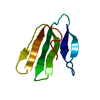
| |||||||||
|---|---|---|---|---|---|---|---|---|---|---|
| 1 |
| |||||||||
| NMR ensembles |
|
-Components
| #1: Protein | Mass: 7293.412 Da / Num. of mol.: 1 / Source method: isolated from a natural source / Source: (natural) Bungarus candidus (cobra) / Secretion: VENOM / References: UniProt: P81782 |
|---|---|
| Has protein modification | Y |
-Experimental details
-Experiment
| Experiment | Method: SOLUTION NMR |
|---|---|
| NMR experiment | Type: 2D NOESY |
| NMR details | Text: The structures were determined using standard 2D homonuclear techniques. |
- Sample preparation
| Details | Contents: 1.7 mM bucandin / Solvent system: 90% H2O/10% D2O |
|---|---|
| Sample conditions | Ionic strength: no salt added / pH: 3 / Pressure: ambient / Temperature: 298 K |
| Crystal grow | *PLUS Method: other / Details: NMR |
-NMR measurement
| Radiation | Protocol: SINGLE WAVELENGTH / Monochromatic (M) / Laue (L): M |
|---|---|
| Radiation wavelength | Relative weight: 1 |
| NMR spectrometer | Type: Bruker AVANCE / Manufacturer: Bruker / Model: AVANCE / Field strength: 600 MHz |
- Processing
| NMR software |
| ||||||||||||||||||||||||||||||||
|---|---|---|---|---|---|---|---|---|---|---|---|---|---|---|---|---|---|---|---|---|---|---|---|---|---|---|---|---|---|---|---|---|---|
| Refinement | Method: distance geometry, simulated annealing, molecular dynamics, torsion angle dynamics Software ordinal: 1 Details: The structures are based on a total of 1363 restraints, 1258 are NOE-derived distance constraints, 61 dihedral angle restraints, 44 distance restraints from hydrogen bonds. | ||||||||||||||||||||||||||||||||
| NMR representative | Selection criteria: closest to the average | ||||||||||||||||||||||||||||||||
| NMR ensemble | Conformer selection criteria: structures with the lowest energy Conformers calculated total number: 6000 / Conformers submitted total number: 20 |
 X-PLOR