 Movie
Movie Controller

[English] 日本語
 Yorodumi
- PDB-1iic: Crystal Structure of Saccharomyces cerevisiae N-myristoyltransfer... -
+ Open data

- Basic information
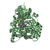
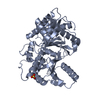
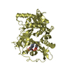
| Entry | Database: PDB / ID: 1iic | ||||||
|---|---|---|---|---|---|---|---|
| Title | Crystal Structure of Saccharomyces cerevisiae N-myristoyltransferase with Bound MyristoylCoA | ||||||
 Components Components | PEPTIDE N-myristoyltransferase | ||||||
 Keywords Keywords | TRANSFERASE | ||||||
| Function / homology |  Function and homology information Function and homology informationInactivation, recovery and regulation of the phototransduction cascade / glycylpeptide N-tetradecanoyltransferase / glycylpeptide N-tetradecanoyltransferase activity / protein localization to membrane / cytosol Similarity search - Function | ||||||
| Biological species |  | ||||||
| Method |  X-RAY DIFFRACTION / MOLECULAR REPLACEMENT / Resolution: 2.2 Å X-RAY DIFFRACTION / MOLECULAR REPLACEMENT / Resolution: 2.2 Å | ||||||
 Authors Authors | Farazi, T.A. / Waksman, G. / Gordon, J.I. | ||||||
 Citation Citation | Journal: Biochemistry / Year: 2001 Title: Structures of Saccharomyces cerevisiae N-myristoyltransferase with bound myristoylCoA and peptide provide insights about substrate recognition and catalysis. Authors: Farazi, T.A. / Waksman, G. / Gordon, J.I. | ||||||
| History |
|
- Structure visualization
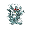
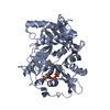
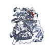
| Structure viewer | Molecule: MolmilJmol/JSmol |
|---|
- Downloads & links
-Download
| PDBx/mmCIF format | 1iic.cif.gz | 188 KB | Display | PDBx/mmCIF format |
|---|---|---|---|---|
| PDB format | pdb1iic.ent.gz | 148.7 KB | Display | PDB format |
| PDBx/mmJSON format | 1iic.json.gz | Tree view | PDBx/mmJSON format | |
| Others |  Other downloads Other downloads |
-Validation report
| Arichive directory | https://data.pdbj.org/pub/pdb/validation_reports/ii/1iicftp://data.pdbj.org/pub/pdb/validation_reports/ii/1iic | HTTPS FTP |
|---|
-Related structure data
| Related structure data |  1iidC  2nmtS S: Starting model for refinement C: citing same article ( |
|---|---|
| Similar structure data |


-Links
 PDBj
PDBj

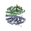
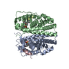
- Assembly
| Deposited unit | 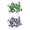
| ||||||||
|---|---|---|---|---|---|---|---|---|---|
| 1 | 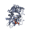
| ||||||||
| 2 | 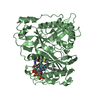
| ||||||||
| Unit cell |
|
-Components
| #1: Protein | Mass: 49013.973 Da / Num. of mol.: 2 Fragment: N-myristoyltransferase (N-terminal 33 residues deleted) Source method: isolated from a genetically manipulated source Source: (gene. exp.) Gene: NMT / Plasmid: pBB501 / Production host:  References: UniProt: P14743, glycylpeptide N-tetradecanoyltransferase #2: Chemical |   Mass: 977.890 Da / Num. of mol.: 2 / Source method: obtained synthetically / Formula: C35H62N7O17P3S Mass: 977.890 Da / Num. of mol.: 2 / Source method: obtained synthetically / Formula: C35H62N7O17P3S#3: Water | ChemComp-HOH / |  Mass: 18.015 Da / Num. of mol.: 266 / Source method: isolated from a natural source / Formula: H2O Mass: 18.015 Da / Num. of mol.: 266 / Source method: isolated from a natural source / Formula: H2O |
|---|
-Experimental details
-Experiment
| Experiment | Method: X-RAY DIFFRACTION / Number of used crystals: 1 |
|---|
- Sample preparation
| Crystal | Density Matthews: 2.5 Å3/Da / Density % sol: 54 % | ||||||||||||||||||||||||||||||
|---|---|---|---|---|---|---|---|---|---|---|---|---|---|---|---|---|---|---|---|---|---|---|---|---|---|---|---|---|---|---|---|
| Crystal grow | Temperature: 293 K / Method: vapor diffusion, hanging drop / pH: 6.4 Details: PEG 4000, ammonium acetate, sodium cacodylate, pH 6.4, VAPOR DIFFUSION, HANGING DROP, temperature 293K | ||||||||||||||||||||||||||||||
| Crystal grow | *PLUS Temperature: 20 ℃ | ||||||||||||||||||||||||||||||
| Components of the solutions | *PLUS
|
-Data collection
| Diffraction | Mean temperature: 103 K |
|---|---|
| Diffraction source | Wavelength: 1.5418 |
| Detector | Type: RIGAKU RAXIS IV / Detector: IMAGE PLATE / Date: Apr 10, 2000 / Details: Yale Mirrors |
| Radiation | Monochromator: graphite / Protocol: SINGLE WAVELENGTH / Monochromatic (M) / Laue (L): M / Scattering type: x-ray |
| Radiation wavelength | Wavelength: 1.5418 Å / Relative weight: 1 |
| Reflection | Resolution: 2.2→30 Å / Num. all: 53396 / Num. obs: 48952 / % possible obs: 91.7 % / Observed criterion σ(F): 2 / Biso Wilson estimate: 20.9 Å2 / Rsym value: 4.8 / Net I/σ(I): 19.4 |
| Reflection shell | Resolution: 2.2→2.28 Å / Mean I/σ(I) obs: 7 / Rsym value: 13.4 / % possible all: 79.59 |
| Reflection | *PLUS Num. measured all: 341253 / Rmerge(I) obs: 0.048 |
- Processing
| Software |
| ||||||||||||||||||||||||||||||||||||||||
|---|---|---|---|---|---|---|---|---|---|---|---|---|---|---|---|---|---|---|---|---|---|---|---|---|---|---|---|---|---|---|---|---|---|---|---|---|---|---|---|---|---|
| Refinement | Method to determine structure: MOLECULAR REPLACEMENT Starting model: PDB ENTRY 2NMT Resolution: 2.2→27.22 Å / Rfactor Rfree error: 0.005 / Data cutoff high absF: 1973354.52 / Data cutoff low absF: 0 / Isotropic thermal model: RESTRAINED / Cross valid method: THROUGHOUT / σ(F): 2 / Stereochemistry target values: Engh & Huber
| ||||||||||||||||||||||||||||||||||||||||
| Solvent computation | Solvent model: FLAT MODEL / Bsol: 37.26 Å2 / ksol: 0.371 e/Å3 | ||||||||||||||||||||||||||||||||||||||||
| Displacement parameters | Biso mean: 28.8 Å2
| ||||||||||||||||||||||||||||||||||||||||
| Refine analyze |
| ||||||||||||||||||||||||||||||||||||||||
| Refinement step | Cycle: LAST / Resolution: 2.2→27.22 Å
| ||||||||||||||||||||||||||||||||||||||||
| Refine LS restraints |
| ||||||||||||||||||||||||||||||||||||||||
| LS refinement shell | Resolution: 2.2→2.34 Å / Rfactor Rfree error: 0.014 / Total num. of bins used: 6
| ||||||||||||||||||||||||||||||||||||||||
| Xplor file |
| ||||||||||||||||||||||||||||||||||||||||
| Software | *PLUS Name: CNS / Version: 0.9 / Classification: refinement | ||||||||||||||||||||||||||||||||||||||||
| Refinement | *PLUS σ(F): 2 / % reflection Rfree: 5.1 % | ||||||||||||||||||||||||||||||||||||||||
| Solvent computation | *PLUS | ||||||||||||||||||||||||||||||||||||||||
| Displacement parameters | *PLUS Biso mean: 28.8 Å2 | ||||||||||||||||||||||||||||||||||||||||
| Refine LS restraints | *PLUS
| ||||||||||||||||||||||||||||||||||||||||
| LS refinement shell | *PLUS Rfactor Rfree: 0.275 / % reflection Rfree: 4.9 % / Rfactor Rwork: 0.232 |
