 Movie
Movie Controller

[English] 日本語
 Yorodumi
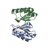
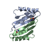
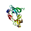
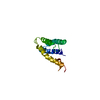
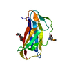
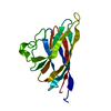
- PDB-1ihw: SOLUTION STRUCTURE OF THE DNA BINDING DOMAIN OF HIV-1 INTEGRASE, ... -
+ Open data

- Basic information
| Entry | Database: PDB / ID: 1ihw | ||||||
|---|---|---|---|---|---|---|---|
| Title | SOLUTION STRUCTURE OF THE DNA BINDING DOMAIN OF HIV-1 INTEGRASE, NMR, 40 STRUCTURES | ||||||
 Components Components | HIV-1 INTEGRASE | ||||||
 Keywords Keywords | DNA BINDING PROTEIN / DNA-BINDING PROTEIN / AIDS / POLYPROTEIN | ||||||
| Function / homology |  Function and homology information Function and homology informationnucleotidyltransferase activity / DNA integration / viral genome integration into host DNA / establishment of integrated proviral latency / Transferases; Transferring phosphorus-containing groups; Nucleotidyltransferases / endonuclease activity / DNA recombination / Hydrolases; Acting on ester bonds / symbiont-mediated suppression of host gene expression / hydrolase activity ...nucleotidyltransferase activity / DNA integration / viral genome integration into host DNA / establishment of integrated proviral latency / Transferases; Transferring phosphorus-containing groups; Nucleotidyltransferases / endonuclease activity / DNA recombination / Hydrolases; Acting on ester bonds / symbiont-mediated suppression of host gene expression / hydrolase activity / symbiont entry into host cell / lipid binding / DNA binding / metal ion binding Similarity search - Function | ||||||
| Biological species |   Human immunodeficiency virus 1 Human immunodeficiency virus 1 | ||||||
| Method | SOLUTION NMR | ||||||
 Authors Authors | Clore, G.M. / Lodi, P.J. / Ernst, J.A. / Gronenborn, A.M. | ||||||
 Citation Citation | Journal: Biochemistry / Year: 1995 Title: Solution structure of the DNA binding domain of HIV-1 integrase. Authors: Lodi, P.J. / Ernst, J.A. / Kuszewski, J. / Hickman, A.B. / Engelman, A. / Craigie, R. / Clore, G.M. / Gronenborn, A.M. | ||||||
| History |
|
- Structure visualization
| Structure viewer | Molecule: MolmilJmol/JSmol |
|---|
- Downloads & links
-Download
| PDBx/mmCIF format | 1ihw.cif.gz | 1.5 MB | Display | PDBx/mmCIF format |
|---|---|---|---|---|
| PDB format | pdb1ihw.ent.gz | 1.3 MB | Display | PDB format |
| PDBx/mmJSON format | 1ihw.json.gz | Tree view | PDBx/mmJSON format | |
| Others |  Other downloads Other downloads |
-Validation report
| Arichive directory | https://data.pdbj.org/pub/pdb/validation_reports/ih/1ihwftp://data.pdbj.org/pub/pdb/validation_reports/ih/1ihw | HTTPS FTP |
|---|
-Related structure data


-Links
 PDBj
PDBj- Assembly
| Deposited unit | 
| |||||||||
|---|---|---|---|---|---|---|---|---|---|---|
| 1 |
| |||||||||
| NMR ensembles |
|
-Components
| #1: Protein | Mass: 6139.186 Da / Num. of mol.: 2 / Fragment: DNA BINDING DOMAIN Source method: isolated from a genetically manipulated source Details: NMR, 40 STRUCTURES / Source: (gene. exp.) Human immunodeficiency virus 1 / Genus: Lentivirus / Production host:  |
|---|
-Experimental details
-Experiment
| Experiment | Method: SOLUTION NMR |
|---|
- Sample preparation
| Crystal grow | *PLUS Method: other / Details: NMR |
|---|
- Processing
| Software |
| ||||||||||||
|---|---|---|---|---|---|---|---|---|---|---|---|---|---|
| NMR software | Name:  X-PLOR / Version: 3.1 / Developer: BRUNGER / Classification: refinement X-PLOR / Version: 3.1 / Developer: BRUNGER / Classification: refinement | ||||||||||||
| Refinement | Software ordinal: 1 Details: THE STRUCTURES WERE CALCULATED USING THE SIMULATED ANNEALING PROTOCOL OF NILGES ET AL. (1988) FEBS LETT. 229, 129 - 136 USING THE PROGRAM XPLOR 3.1 (BRUNGER) MODIFIED TO INCORPORATE COUPLING ...Details: THE STRUCTURES WERE CALCULATED USING THE SIMULATED ANNEALING PROTOCOL OF NILGES ET AL. (1988) FEBS LETT. 229, 129 - 136 USING THE PROGRAM XPLOR 3.1 (BRUNGER) MODIFIED TO INCORPORATE COUPLING CONSTANT (GARRETT ET AL. (1984) J. MAGN RESON. SERIES B 104, 99 - 103), CARBON CHEMICAL SHIFT (KUSZEWSKI ET AL. (1995) J. MAGN. RESON. SERIES B 106, 92 - 96) AND 1H CHEMICAL SHIFT (KUSZEWSKI ET AL., 1995 J. MAGN RESON. SERIES B IN PRESS) RESTRAINTS THE 3D STRUCTURE OF THE HUMAN SRY-DNA COMPLEX SOLVED BY MULTI-DIMENSIONAL HETERONUCLEAR-EDITED AND -FILTERED NMR IS BASED ON 2386 EXPERIMENTAL RESTRAINTS (FOR THE DIMER): (A) INTRASUBUNIT: 332 SEQUENTIAL (|I-J|=1), 202 MEDIUM RANGE (1 < |I-J| >=5) AND 530 LONG RANGE (|I-J| >5) INTERRESIDUES 318 INTRARESIDUE APPROXIMATE INTERPROTON DISTANCE RESTRAINTS; 74 DISTANCE RESTRAINTS FOR 37 HYDROGEN BONDS; 192 TORSION ANGLE (98 PHI, 20 PSI, 50 CHI1 AND 24 CHI2) RESTRAINTS; 78 THREE-BOND HN-HA COUPLING CONSTANT RESTRAINTS; 194 (100 CALPHA AND 94 CBETA) 13C SHIFT RESTRAINTS; AND 392 1H CHEMICAL SHIFT RESTRAINTS (102 CAH, 52 METHYL AND 238 OTHERS). (B) 44 INTERSUBUNIT INTERPROTON DISTANCE RESTRAINTS (C) 30 AMBIGUOUS INTERPROTON DISTANCE RESTRAINTS THAT CAN ARISE FROM INTRA AND/OR INTERSUBUNIT INTERACTIONS. THE STRUCTURE FOUND IN PDB ENTRY 1IHV IS THE RESTRAINED REGULARIZED MEAN STRUCTURE AND THE LAST COLUMN REPRESENTS THE RMS OF THE 40 INDIVIDUAL SIMULATED ANNEALING STRUCTURES FOUND IN THIS ENTRY ABOUT THE MEAN COORDINATE POSITIONS. THE LAST COLUMN IN THE INDIVIDUAL SA STRUCTURES HAS NO MEANING. | ||||||||||||
| NMR ensemble | Conformers submitted total number: 40 |
