 Movie
Movie Controller


+ Open data

- Basic information
| Entry | Database: PDB / ID: 1ihg | ||||||
|---|---|---|---|---|---|---|---|
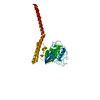
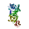
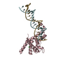
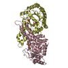
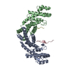
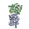
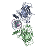
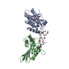
| Title | Bovine Cyclophilin 40, monoclinic form | ||||||
 Components Components | Cyclophilin 40 | ||||||
 Keywords Keywords | ISOMERASE / PPIASE IMMUNOPHILIN TETRATRICOPEPTIDE | ||||||
| Function / homology |  Function and homology information Function and homology informationcellular response to UV-A / lipid droplet organization / cyclosporin A binding / transcription factor binding / positive regulation of viral genome replication / Hsp70 protein binding / nuclear estrogen receptor binding / peptidylprolyl isomerase / positive regulation of protein secretion / peptidyl-prolyl cis-trans isomerase activity ...cellular response to UV-A / lipid droplet organization / cyclosporin A binding / transcription factor binding / positive regulation of viral genome replication / Hsp70 protein binding / nuclear estrogen receptor binding / peptidylprolyl isomerase / positive regulation of protein secretion / peptidyl-prolyl cis-trans isomerase activity / Hsp90 protein binding / protein transport / protein folding / protein-containing complex assembly / cilium / positive regulation of apoptotic process / apoptotic process / nucleolus / negative regulation of transcription by RNA polymerase II / nucleoplasm / nucleus / cytoplasm / cytosol Similarity search - Function | ||||||
| Biological species |  | ||||||
| Method |  X-RAY DIFFRACTION / SYNCHROTRON / MOLECULAR REPLACEMENT / Resolution: 1.8 Å X-RAY DIFFRACTION / SYNCHROTRON / MOLECULAR REPLACEMENT / Resolution: 1.8 Å | ||||||
 Authors Authors | Taylor, P. / Dornan, J. / Carrello, A. / Minchin, R.F. / Ratajczak, T. / Walkinshaw, M.D. | ||||||
 Citation Citation | Journal: Structure / Year: 2001 Title: Two structures of cyclophilin 40: folding and fidelity in the TPR domains. Authors: Taylor, P. / Dornan, J. / Carrello, A. / Minchin, R.F. / Ratajczak, T. / Walkinshaw, M.D. | ||||||
| History |
|
- Structure visualization
| Structure viewer | Molecule: MolmilJmol/JSmol |
|---|
- Downloads & links
-Download
| PDBx/mmCIF format | 1ihg.cif.gz | 92.3 KB | Display | PDBx/mmCIF format |
|---|---|---|---|---|
| PDB format | pdb1ihg.ent.gz | 69.3 KB | Display | PDB format |
| PDBx/mmJSON format | 1ihg.json.gz | Tree view | PDBx/mmJSON format | |
| Others |  Other downloads Other downloads |
-Validation report
| Arichive directory | https://data.pdbj.org/pub/pdb/validation_reports/ih/1ihgftp://data.pdbj.org/pub/pdb/validation_reports/ih/1ihg | HTTPS FTP |
|---|
-Related structure data

-Links
 PDBj
PDBj


- Assembly
| Deposited unit | 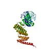
| ||||||||
|---|---|---|---|---|---|---|---|---|---|
| 1 |
| ||||||||
| Unit cell |
|
-Components
| #1: Protein | Mass: 40678.316 Da / Num. of mol.: 1 Source method: isolated from a genetically manipulated source Source: (gene. exp.)  |
|---|---|
| #2: Chemical | ChemComp-GOL /   Mass: 92.094 Da / Num. of mol.: 1 / Source method: obtained synthetically / Formula: C3H8O3 Mass: 92.094 Da / Num. of mol.: 1 / Source method: obtained synthetically / Formula: C3H8O3 |
| #3: Water | ChemComp-HOH /  Mass: 18.015 Da / Num. of mol.: 489 / Source method: isolated from a natural source / Formula: H2O Mass: 18.015 Da / Num. of mol.: 489 / Source method: isolated from a natural source / Formula: H2O |
-Experimental details
-Experiment
| Experiment | Method: X-RAY DIFFRACTION / Number of used crystals: 1 |
|---|
- Sample preparation
| Crystal | Density Matthews: 2 Å3/Da / Density % sol: 54.99 % | |||||||||||||||||||||||||||||||||||||||||||||||||||||||||||||||
|---|---|---|---|---|---|---|---|---|---|---|---|---|---|---|---|---|---|---|---|---|---|---|---|---|---|---|---|---|---|---|---|---|---|---|---|---|---|---|---|---|---|---|---|---|---|---|---|---|---|---|---|---|---|---|---|---|---|---|---|---|---|---|---|---|
| Crystal grow | Temperature: 277 K / Method: vapor diffusion, hanging drop / pH: 6.1 Details: PEG 4000, MES Glycerol Sodium Chloride, pH 6.1, VAPOR DIFFUSION, HANGING DROP, temperature 277K | |||||||||||||||||||||||||||||||||||||||||||||||||||||||||||||||
| Crystal grow | *PLUS Temperature: 4 ℃ / pH: 8 | |||||||||||||||||||||||||||||||||||||||||||||||||||||||||||||||
| Components of the solutions | *PLUS
|
-Data collection
| Diffraction | Mean temperature: 100 K |
|---|---|
| Diffraction source | Source: SYNCHROTRON / Site: SRS  / Beamline: PX9.6 / Wavelength: 0.87 / Wavelength: 0.87 Å / Beamline: PX9.6 / Wavelength: 0.87 / Wavelength: 0.87 Å |
| Detector | Type: ADSC QUANTUM 4 / Detector: CCD / Date: Jun 10, 1999 |
| Radiation | Monochromator: Single Crystal Si / Protocol: SINGLE WAVELENGTH / Monochromatic (M) / Laue (L): M / Scattering type: x-ray |
| Radiation wavelength | Wavelength: 0.87 Å / Relative weight: 1 |
| Reflection | Resolution: 1.8→28 Å / Num. all: 41259 / Num. obs: 41259 / % possible obs: 98.3 % / Observed criterion σ(F): 0 / Observed criterion σ(I): 0 / Redundancy: 3.7 % / Biso Wilson estimate: 13.448 Å2 / Rmerge(I) obs: 0.05 / Net I/σ(I): 20.67 |
| Reflection shell | Resolution: 1.8→1.83 Å / Redundancy: 1.87 % / Rmerge(I) obs: 0.096 / Mean I/σ(I) obs: 9.03 / Num. unique all: 1769 / % possible all: 87.3 |
| Reflection | *PLUS Rmerge(I) obs: 0.05 |
- Processing
| Software |
| |||||||||||||||||||||||||||||||||
|---|---|---|---|---|---|---|---|---|---|---|---|---|---|---|---|---|---|---|---|---|---|---|---|---|---|---|---|---|---|---|---|---|---|---|
| Refinement | Method to determine structure: MOLECULAR REPLACEMENT Starting model: CYCLOPHILIN A Resolution: 1.8→24 Å / Num. parameters: 13223 / Num. restraintsaints: 11467 / Cross valid method: FREE R / σ(F): 0 / σ(I): 0 / Stereochemistry target values: ENGH AND HUBER
| |||||||||||||||||||||||||||||||||
| Refine analyze | Occupancy sum hydrogen: 0 / Occupancy sum non hydrogen: 3305 | |||||||||||||||||||||||||||||||||
| Refinement step | Cycle: LAST / Resolution: 1.8→24 Å
| |||||||||||||||||||||||||||||||||
| Refine LS restraints |
| |||||||||||||||||||||||||||||||||
| Software | *PLUS Name: SHELXL-97 / Classification: refinement | |||||||||||||||||||||||||||||||||
| Refinement | *PLUS σ(F): 0 / % reflection Rfree: 5 % | |||||||||||||||||||||||||||||||||
| Solvent computation | *PLUS | |||||||||||||||||||||||||||||||||
| Displacement parameters | *PLUS | |||||||||||||||||||||||||||||||||
| Refine LS restraints | *PLUS
|
