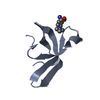
Mass: 425.501 Da / Num. of mol.: 1 / Source method: obtained synthetically / Formula: C22H23N3O4S
-
Experimental details
-
Experiment
Experiment
Method: SOLUTION NMR
NMR experiment
Conditions-ID
Experiment-ID
Solution-ID
Type
1
1
1
HNHA
1
2
1
HNHB
1
3
1
3D-13C/15N-NOESY
1
4
1
HCCHTOCSY
1
5
1
3D 15N-separated NOESY
1
6
1
HN(CA)CB
NMR details
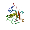
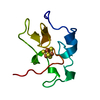
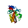
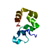
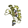
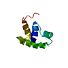
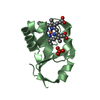
Text: The structure of the protein was determined using multidimensional NMR spectroscopy. The structure of EMD 57033 in the complex was determined using 15N/13C-filtered-NOESY and DIPSI experiments. ...Text: The structure of the protein was determined using multidimensional NMR spectroscopy. The structure of EMD 57033 in the complex was determined using 15N/13C-filtered-NOESY and DIPSI experiments. The contacts between protein and EMD 57033 were determined using 15N/13C-filtered/edited experiments.
Method: simulated annealing / Software ordinal: 1 Details: ~1000 NOE restraints were used. 61 dihedral restraints as well as 14 NOE restraints between drug and protein.
NMR representative
Selection criteria: closest to the minimized average structure
NMR ensemble
Conformer selection criteria: all calculated structures submitted Conformers calculated total number: 30 / Conformers submitted total number: 30
+
About Yorodumi
-
News
-
Feb 9, 2022. New format data for meta-information of EMDB entries
New format data for meta-information of EMDB entries
Version 3 of the EMDB header file is now the official format.
The previous official version 1.9 will be removed from the archive.
In the structure databanks used in Yorodumi, some data are registered as the other names, "COVID-19 virus" and "2019-nCoV". Here are the details of the virus and the list of structure data.
Jan 31, 2019. EMDB accession codes are about to change! (news from PDBe EMDB page)
EMDB accession codes are about to change! (news from PDBe EMDB page)
The allocation of 4 digits for EMDB accession codes will soon come to an end. Whilst these codes will remain in use, new EMDB accession codes will include an additional digit and will expand incrementally as the available range of codes is exhausted. The current 4-digit format prefixed with “EMD-” (i.e. EMD-XXXX) will advance to a 5-digit format (i.e. EMD-XXXXX), and so on. It is currently estimated that the 4-digit codes will be depleted around Spring 2019, at which point the 5-digit format will come into force.
The EM Navigator/Yorodumi systems omit the EMD- prefix.
Related info.:Q: What is EMD? / ID/Accession-code notation in Yorodumi/EM Navigator
Yorodumi is a browser for structure data from EMDB, PDB, SASBDB, etc.
This page is also the successor to EM Navigator detail page, and also detail information page/front-end page for Omokage search.
The word "yorodu" (or yorozu) is an old Japanese word meaning "ten thousand". "mi" (miru) is to see.
Related info.:EMDB / PDB / SASBDB / Comparison of 3 databanks / Yorodumi Search / Aug 31, 2016. New EM Navigator & Yorodumi / Yorodumi Papers / Jmol/JSmol / Function and homology information / Changes in new EM Navigator and Yorodumi
 Movie
Movie Controller

 Yorodumi
 Open data
 Basic information
 Components
 Keywords
 Function and homology information
 Homo sapiens (human)
 Authors
 Citation
 Structure visualization
 Downloads & links
 Other downloads


 PDBj
PDBj


 Assembly
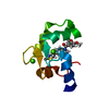


 Mass: 40.078 Da / Num. of mol.: 2 / Source method: obtained synthetically / Formula: Ca
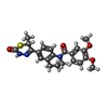
 Mass: 425.501 Da / Num. of mol.: 1 / Source method: obtained synthetically / Formula: C22H23N3O4S
 Sample preparation
 Processing
 X-PLOR / Version: 3.851 / Developer: Brunger, A. / Classification: refinement