 Movie
Movie Controller


+ Open data

- Basic information
| Entry | Database: PDB / ID: 1igy | |||||||||
|---|---|---|---|---|---|---|---|---|---|---|
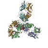
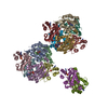
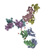
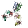
| Title | STRUCTURE OF IMMUNOGLOBULIN | |||||||||
 Components Components | (IGG1 INTACT ANTIBODY MAB61.1.3) x 2 | |||||||||
 Keywords Keywords | IMMUNOGLOBULIN / INTACT IMMUNOGLOBULIN / V REGION / C REGION / HINGE REGION | |||||||||
| Function / homology |  Function and homology information Function and homology informationClassical antibody-mediated complement activation / FCGR activation / Role of phospholipids in phagocytosis / Regulation of Complement cascade / positive regulation of type IIa hypersensitivity / humoral immune response mediated by circulating immunoglobulin / phagocytosis, recognition / Regulation of actin dynamics for phagocytic cup formation / positive regulation of type I hypersensitivity / antibody-dependent cellular cytotoxicity ...Classical antibody-mediated complement activation / FCGR activation / Role of phospholipids in phagocytosis / Regulation of Complement cascade / positive regulation of type IIa hypersensitivity / humoral immune response mediated by circulating immunoglobulin / phagocytosis, recognition / Regulation of actin dynamics for phagocytic cup formation / positive regulation of type I hypersensitivity / antibody-dependent cellular cytotoxicity / Fc-gamma receptor I complex binding / alpha-beta T cell receptor complex / immunoglobulin complex, circulating / phagocytosis, engulfment / immunoglobulin receptor binding / IgG immunoglobulin complex / immunoglobulin mediated immune response / complement activation, classical pathway / antigen binding / B cell differentiation / positive regulation of phagocytosis / positive regulation of immune response / antibacterial humoral response / adaptive immune response / defense response to bacterium / external side of plasma membrane / : / extracellular region / plasma membrane / cytoplasm Similarity search - Function | |||||||||
| Biological species |  | |||||||||
| Method |  X-RAY DIFFRACTION / SYNCHROTRON / MOLECULAR REPLACEMENT / Resolution: 3.2 Å X-RAY DIFFRACTION / SYNCHROTRON / MOLECULAR REPLACEMENT / Resolution: 3.2 Å | |||||||||
 Authors Authors | Harris, L.J. / McPherson, A. | |||||||||
 Citation Citation | Journal: J.Mol.Biol. / Year: 1998 Title: Crystallographic structure of an intact IgG1 monoclonal antibody. Authors: Harris, L.J. / Skaletsky, E. / McPherson, A. #1: Journal: Proteins / Year: 1995Title: Crystallization of Intact Monoclonal Antibodies Authors: Harris, L.J. / Skaletsky, E. / McPherson, A. | |||||||||
| History |
|
- Structure visualization
| Structure viewer | Molecule: MolmilJmol/JSmol |
|---|
- Downloads & links
-Download
| PDBx/mmCIF format | 1igy.cif.gz | 297.6 KB | Display | PDBx/mmCIF format |
|---|---|---|---|---|
| PDB format | pdb1igy.ent.gz | 239.9 KB | Display | PDB format |
| PDBx/mmJSON format | 1igy.json.gz | Tree view | PDBx/mmJSON format | |
| Others |  Other downloads Other downloads |
-Validation report
| Arichive directory | https://data.pdbj.org/pub/pdb/validation_reports/ig/1igyftp://data.pdbj.org/pub/pdb/validation_reports/ig/1igy | HTTPS FTP |
|---|
-Related structure data
| Related structure data | 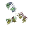 1igtS  1mcpS  2hfl S: Starting model for refinement |
|---|---|
| Similar structure data |


-Links
 PDBj
PDBj


- Assembly
| Deposited unit | 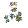
| ||||||||
|---|---|---|---|---|---|---|---|---|---|
| 1 |
| ||||||||
| Unit cell |
| ||||||||
| Details | THERE IS ONE ENTIRE ANTIBODY MOLECULE PER ASYMMETRIC UNIT. |
-Components
| #1: Antibody | Mass: 23519.973 Da / Num. of mol.: 2 / Source method: isolated from a natural source Details: HYBRIDOMA MONOCLONAL ANTIBODY, AGAINST PHENOBARBITAL Source: (natural) #2: Antibody | Mass: 47906.895 Da / Num. of mol.: 2 / Source method: isolated from a natural source Details: HYBRIDOMA MONOCLONAL ANTIBODY, AGAINST PHENOBARBITAL Source: (natural) #3: Polysaccharide | beta-D-galactopyranose-(1-4)-2-acetamido-2-deoxy-beta-D-glucopyranose-(1-2)-alpha-D-mannopyranose- ...beta-D-galactopyranose-(1-4)-2-acetamido-2-deoxy-beta-D-glucopyranose-(1-2)-alpha-D-mannopyranose-(1-6)-[2-acetamido-2-deoxy-beta-D-glucopyranose-(1-2)-alpha-D-mannopyranose-(1-3)]beta-D-mannopyranose-(1-4)-2-acetamido-2-deoxy-alpha-D-glucopyranose-(1-4)-[beta-L-fucopyranose-(1-6)]2-acetamido-2-deoxy-beta-D-glucopyranose | Source method: isolated from a genetically manipulated source #4: Polysaccharide | beta-D-galactopyranose-(1-4)-2-acetamido-2-deoxy-beta-D-glucopyranose-(1-2)-alpha-D-mannopyranose- ...beta-D-galactopyranose-(1-4)-2-acetamido-2-deoxy-beta-D-glucopyranose-(1-2)-alpha-D-mannopyranose-(1-6)-[2-acetamido-2-deoxy-beta-D-glucopyranose-(1-2)-alpha-D-mannopyranose-(1-3)]beta-D-mannopyranose-(1-4)-2-acetamido-2-deoxy-alpha-D-glucopyranose-(1-4)-[alpha-L-fucopyranose-(1-6)]2-acetamido-2-deoxy-beta-D-glucopyranose | Source method: isolated from a genetically manipulated source Has protein modification | Y | Sequence details | THE INTACT ANTIBODY IS NUMBERED ACCORDING TO THE CONVENTION OF E. KABAT [KABAT ET AL. (1991) ...THE INTACT ANTIBODY IS NUMBERED ACCORDING TO THE CONVENTION | |
|---|
-Experimental details
-Experiment
| Experiment | Method: X-RAY DIFFRACTION / Number of used crystals: 5 |
|---|
- Sample preparation
| Crystal | Density Matthews: 3 Å3/Da / Density % sol: 59 % |
|---|---|
| Crystal grow | pH: 5.1 Details: 4 MICROLITERS OF 8.7 MG/ML MAB 61.1.3, 2 MICROLITERS OF 50 MILLIMOLAR SODIUM CITRATE PH 5, 1 MICROLITER OF N-TRIDECYL-B-D-MALTOSIDE, AND 5 MICROLITERS OF 12% PEG 3350 EQUILIBRATED AGAINST ...Details: 4 MICROLITERS OF 8.7 MG/ML MAB 61.1.3, 2 MICROLITERS OF 50 MILLIMOLAR SODIUM CITRATE PH 5, 1 MICROLITER OF N-TRIDECYL-B-D-MALTOSIDE, AND 5 MICROLITERS OF 12% PEG 3350 EQUILIBRATED AGAINST 700 MICROLITERS OF 12% PEG 3350, AT ROOM TEMPERATURE (CRYSCHEM PLATE)., pH 5.1 Temp details: room temp |
| Crystal | *PLUS |
| Crystal grow | *PLUS Method: vapor diffusion, sitting dropDetails: Harris, L.J., (1995) Proteins: Struct.,Funct., Genet., 23, 285. PH range low: 5 / PH range high: 4.5 |
| Components of the solutions | *PLUS Conc.: 12 %(w/v) / Common name: PEG3350 |
-Data collection
| Diffraction | Mean temperature: 291 K |
|---|---|
| Diffraction source | Source: SYNCHROTRON / Site: NSLS  / Beamline: X12C / Wavelength: 1.15 / Beamline: X12C / Wavelength: 1.15 |
| Detector | Type: MARRESEARCH / Detector: IMAGE PLATE / Date: Jun 1, 1995 / Details: MIRROR |
| Radiation | Monochromator: CRYSTAL TYPE SI(111) / Monochromatic (M) / Laue (L): M / Scattering type: x-ray |
| Radiation wavelength | Wavelength: 1.15 Å / Relative weight: 1 |
| Reflection | Resolution: 3.2→99 Å / Num. obs: 27971 / % possible obs: 95.6 % / Redundancy: 3.3 % / Biso Wilson estimate: 45.2 Å2 / Rmerge(I) obs: 0.145 / Net I/σ(I): 6.4 |
- Processing
| Software |
| ||||||||||||||||||||||||||||||||||||||||||||||||||||||||||||
|---|---|---|---|---|---|---|---|---|---|---|---|---|---|---|---|---|---|---|---|---|---|---|---|---|---|---|---|---|---|---|---|---|---|---|---|---|---|---|---|---|---|---|---|---|---|---|---|---|---|---|---|---|---|---|---|---|---|---|---|---|---|
| Refinement | Method to determine structure: MOLECULAR REPLACEMENT Starting model: VL:VH DOMAIN PAIR OF PDB ENTRY 1MCP, CL:CH1 DOMAIN PAIR OF PDB ENTRY 2HFL, FC FRAGMENT OF PDB ENTRY 1IGT Resolution: 3.2→20 Å / Rfactor Rfree error: 0.007 / Data cutoff high absF: 1000000 / Data cutoff low absF: 0.001 / Isotropic thermal model: GROUP / Cross valid method: THROUGHOUT / σ(F): 3 Details: BULK SOLVENT MODEL USED. THE LOWER HINGE REGION OF CHAIN B WAS DISORDERED BUT MODELED STEREOCHEMICALLY.
| ||||||||||||||||||||||||||||||||||||||||||||||||||||||||||||
| Displacement parameters | Biso mean: 57.1 Å2 | ||||||||||||||||||||||||||||||||||||||||||||||||||||||||||||
| Refine analyze |
| ||||||||||||||||||||||||||||||||||||||||||||||||||||||||||||
| Refinement step | Cycle: LAST / Resolution: 3.2→20 Å
| ||||||||||||||||||||||||||||||||||||||||||||||||||||||||||||
| Refine LS restraints |
| ||||||||||||||||||||||||||||||||||||||||||||||||||||||||||||
| Refine LS restraints NCS | NCS model details: RESTRAINED | ||||||||||||||||||||||||||||||||||||||||||||||||||||||||||||
| LS refinement shell | Resolution: 3.2→3.4 Å / Rfactor Rfree error: 0.03 / Total num. of bins used: 6
| ||||||||||||||||||||||||||||||||||||||||||||||||||||||||||||
| Xplor file |
| ||||||||||||||||||||||||||||||||||||||||||||||||||||||||||||
| Software | *PLUS Name: X-PLOR / Version: 3.8 / Classification: refinement | ||||||||||||||||||||||||||||||||||||||||||||||||||||||||||||
| Refinement | *PLUS Num. reflection obs: 19081 | ||||||||||||||||||||||||||||||||||||||||||||||||||||||||||||
| Solvent computation | *PLUS | ||||||||||||||||||||||||||||||||||||||||||||||||||||||||||||
| Displacement parameters | *PLUS | ||||||||||||||||||||||||||||||||||||||||||||||||||||||||||||
| Refine LS restraints | *PLUS
|
