 Movie
Movie Controller

[English] 日本語
 Yorodumi
- PDB-1igw: Crystal Structure of the Isocitrate Lyase from the A219C mutant o... -
+ Open data

- Basic information
| Entry | Database: PDB / ID: 1igw | |||||||||
|---|---|---|---|---|---|---|---|---|---|---|
| Title | Crystal Structure of the Isocitrate Lyase from the A219C mutant of Escherichia coli | |||||||||
 Components Components | Isocitrate lyase | |||||||||
 Keywords Keywords | LYASE / BETA BARREL | |||||||||
| Function / homology |  Function and homology information Function and homology informationisocitrate lyase / cation binding / isocitrate lyase activity / glyoxylate cycle / tricarboxylic acid cycle / metal ion binding / cytosol Similarity search - Function | |||||||||
| Biological species |  | |||||||||
| Method |  X-RAY DIFFRACTION / SYNCHROTRON / MOLECULAR REPLACEMENT / Resolution: 2.1 Å X-RAY DIFFRACTION / SYNCHROTRON / MOLECULAR REPLACEMENT / Resolution: 2.1 Å | |||||||||
 Authors Authors | Britton, K.L. / Abeysinghe, I.S.B. / Baker, P.J. / Barynin, V. / Diehl, P. / Langridge, S.J. / McFadden, B.A. / Sedelnikova, S.E. / Stillman, T.J. / Weeradechapon, K. / Rice, D.W. | |||||||||
 Citation Citation | Journal: Acta Crystallogr.,Sect.D / Year: 2001 Title: The structure and domain organization of Escherichia coli isocitrate lyase. Authors: Britton, K.L. / Abeysinghe, I.S. / Baker, P.J. / Barynin, V. / Diehl, P. / Langridge, S.J. / McFadden, B.A. / Sedelnikova, S.E. / Stillman, T.J. / Weeradechapon, K. / Rice, D.W. #1: Journal: J.Mol.Biol. / Year: 1991Title: Use of Chemical Modification in the Crystallization of Isocitrate Lyase from Escherichia coli Authors: Abeysinghe, S.I. / Baker, P.J. / Rice, D.W. / Rodgers, H.F. / Stillman, T.J. / Ko, Y.H. / McFadden, B.A. / Nimmo, H.G. | |||||||||
| History |
|
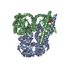
- Structure visualization
| Structure viewer | Molecule: MolmilJmol/JSmol |
|---|
- Downloads & links
-Download
| PDBx/mmCIF format | 1igw.cif.gz | 340.2 KB | Display | PDBx/mmCIF format |
|---|---|---|---|---|
| PDB format | pdb1igw.ent.gz | 274.1 KB | Display | PDB format |
| PDBx/mmJSON format | 1igw.json.gz | Tree view | PDBx/mmJSON format | |
| Others |  Other downloads Other downloads |
-Validation report
| Arichive directory | https://data.pdbj.org/pub/pdb/validation_reports/ig/1igwftp://data.pdbj.org/pub/pdb/validation_reports/ig/1igw | HTTPS FTP |
|---|
-Related structure data
| Similar structure data |
|---|


-Links
 PDBj
PDBj


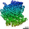
- Assembly
| Deposited unit | 
| ||||||||
|---|---|---|---|---|---|---|---|---|---|
| 1 |
| ||||||||
| Unit cell |
| ||||||||
| Details | The given tetramer defines the biological assembly |
-Components
| #1: Protein | Mass: 47598.395 Da / Num. of mol.: 4 / Mutation: A219C Source method: isolated from a genetically manipulated source Source: (gene. exp.) #2: Chemical | ChemComp-HG /   Mass: 200.590 Da / Num. of mol.: 21 / Source method: obtained synthetically / Formula: Hg Mass: 200.590 Da / Num. of mol.: 21 / Source method: obtained synthetically / Formula: Hg#3: Chemical | ChemComp-MG /   Mass: 24.305 Da / Num. of mol.: 4 / Source method: obtained synthetically / Formula: Mg Mass: 24.305 Da / Num. of mol.: 4 / Source method: obtained synthetically / Formula: Mg#4: Chemical | ChemComp-PYR /   Mass: 88.062 Da / Num. of mol.: 4 / Source method: obtained synthetically / Formula: C3H4O3 Mass: 88.062 Da / Num. of mol.: 4 / Source method: obtained synthetically / Formula: C3H4O3#5: Water | ChemComp-HOH / |  Mass: 18.015 Da / Num. of mol.: 767 / Source method: isolated from a natural source / Formula: H2O Mass: 18.015 Da / Num. of mol.: 767 / Source method: isolated from a natural source / Formula: H2O |
|---|
-Experimental details
-Experiment
| Experiment | Method: X-RAY DIFFRACTION / Number of used crystals: 1 |
|---|
- Sample preparation
| Crystal | Density Matthews: 2.38 Å3/Da / Density % sol: 48.21 % | ||||||||||||||||||||||||||||||
|---|---|---|---|---|---|---|---|---|---|---|---|---|---|---|---|---|---|---|---|---|---|---|---|---|---|---|---|---|---|---|---|
| Crystal grow | Temperature: 290 K / Method: vapor diffusion, hanging drop / pH: 7 Details: MEPEG 2000, magnesium chloride, ethyl mercury thiosalicylate, HEPES, pH 7.0, VAPOR DIFFUSION, HANGING DROP, temperature 290K | ||||||||||||||||||||||||||||||
| Crystal grow | *PLUS | ||||||||||||||||||||||||||||||
| Components of the solutions | *PLUS
|
-Data collection
| Diffraction | Mean temperature: 298 K |
|---|---|
| Diffraction source | Source: SYNCHROTRON / Site: SRS  / Beamline: PX9.5 / Wavelength: 0.92 Å / Beamline: PX9.5 / Wavelength: 0.92 Å |
| Detector | Type: MARRESEARCH / Detector: IMAGE PLATE / Date: Nov 19, 1995 / Details: mirrors |
| Radiation | Protocol: SINGLE WAVELENGTH / Monochromatic (M) / Laue (L): M / Scattering type: x-ray |
| Radiation wavelength | Wavelength: 0.92 Å / Relative weight: 1 |
| Reflection | Resolution: 2.1→33.3 Å / Num. all: 188246 / Num. obs: 96329 / % possible obs: 94.1 % / Redundancy: 2 % / Rmerge(I) obs: 0.051 |
| Reflection shell | Resolution: 2.1→2.15 Å / Redundancy: 1.9 % / Rmerge(I) obs: 0.216 / Num. unique all: 6893 / % possible all: 91.3 |
| Reflection | *PLUS Num. measured all: 188246 |
| Reflection shell | *PLUS % possible obs: 91.3 % / Num. unique obs: 6893 / Num. measured obs: 13435 |
- Processing
| Software |
| |||||||||||||||||||||||||||||||||
|---|---|---|---|---|---|---|---|---|---|---|---|---|---|---|---|---|---|---|---|---|---|---|---|---|---|---|---|---|---|---|---|---|---|---|
| Refinement | Method to determine structure: MOLECULAR REPLACEMENT / Resolution: 2.1→15 Å / Stereochemistry target values: Engh and Huber
| |||||||||||||||||||||||||||||||||
| Displacement parameters | Biso mean: 31 Å2 | |||||||||||||||||||||||||||||||||
| Refinement step | Cycle: LAST / Resolution: 2.1→15 Å
| |||||||||||||||||||||||||||||||||
| Refine LS restraints |
| |||||||||||||||||||||||||||||||||
| Software | *PLUS Name: REFMAC / Classification: refinement | |||||||||||||||||||||||||||||||||
| Refinement | *PLUS Highest resolution: 2.1 Å / Rfactor obs: 0.184 | |||||||||||||||||||||||||||||||||
| Solvent computation | *PLUS | |||||||||||||||||||||||||||||||||
| Displacement parameters | *PLUS Biso mean: 31 Å2 | |||||||||||||||||||||||||||||||||
| Refine LS restraints | *PLUS
| |||||||||||||||||||||||||||||||||
| LS refinement shell | *PLUS Rfactor Rfree: 0.297 / Rfactor obs: 0.234 |
