 Movie
Movie Controller

[English] 日本語
 Yorodumi
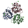
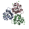
- PDB-1idp: Crystal structure of scytalone dehydratase F162A mutant in the un... -
+ Open data

- Basic information
| Entry | Database: PDB / ID: 1idp | ||||||
|---|---|---|---|---|---|---|---|
| Title | Crystal structure of scytalone dehydratase F162A mutant in the unligated state | ||||||
 Components Components | SCYTALONE DEHYDRATASE | ||||||
 Keywords Keywords | LYASE / MELANINE BIOSYNTHESIS | ||||||
| Function / homology |  Function and homology information Function and homology informationscytalone dehydratase / scytalone dehydratase activity / melanin biosynthetic process / endosome / metal ion binding Similarity search - Function | ||||||
| Biological species |  Magnaporthe grisea (fungus) Magnaporthe grisea (fungus) | ||||||
| Method |  X-RAY DIFFRACTION / SYNCHROTRON / MOLECULAR REPLACEMENT / Resolution: 1.45 Å X-RAY DIFFRACTION / SYNCHROTRON / MOLECULAR REPLACEMENT / Resolution: 1.45 Å | ||||||
 Authors Authors | Nakasako, M. / Motoyama, T. / Yamaguchi, I. | ||||||
 Citation Citation | Journal: Acta Crystallogr.,Sect.D / Year: 2002 Title: Crystallization of scytalone dehydratase F162A mutant in the unligated state and a preliminary X-ray diffraction study at 37 K Authors: Motoyama, T. / Nakasako, M. / Yamaguchi, I. #1: Journal: Biochemistry / Year: 1998Title: Cryogenic X-ray crystal structure analysis for the complex of scytalone dehydratase of a rice blast fungus and its tight-binding inhibitor, carpropamid: the structural basis of tight-binding inhibition Authors: Nakasako, M. / Motoyama, T. / Kurahashi, Y. / Yamaguchi, I. #2: Journal: Proteins / Year: 1999Title: High-resolution structures of scytalone dehydratase-inhibitor complexes crystallized at physiological pH Authors: Wawrzak, Z. / Sandalova, T. / Steffens, J.J. / Basarab, G.S. / Lundqvist, T. / Lindqvist, Y. / Jordan, D.B. #3: Journal: Structure / Year: 1994Title: Crystal structure of scytalone dehydratase--a disease determinant of the rice pathogen, Magnaporthe grisea Authors: Lundqvist, T. / Rice, J. / Hodge, C.N. / Basarab, G.S. / Pierce, J. / Lindqvist, Y. #4: Journal: Biochemistry / Year: 1998Title: Structure-based design of potent inhibitors of scytalone dehydratase: displacement of a water molecule from the active site Authors: Chen, J.M. / Xu, S.L. / Wawrzak, Z. / Basarab, G.S. / Jordan, D.B. #5: Journal: Biosci.Biotechnol.Biochem. / Year: 1998 Title: cDNA cloning, expression, and mutagenesis of scytalone dehydratase needed for pathogenicity of the rice blast fungus, Pyricularia oryzae Authors: Motoyama, T. / Imanishi, K. / Yamaguchi, I. | ||||||
| History |
|
- Structure visualization
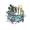
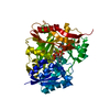
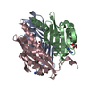
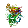
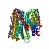
| Structure viewer | Molecule: MolmilJmol/JSmol |
|---|
- Downloads & links
-Download
| PDBx/mmCIF format | 1idp.cif.gz | 100.1 KB | Display | PDBx/mmCIF format |
|---|---|---|---|---|
| PDB format | pdb1idp.ent.gz | 77.1 KB | Display | PDB format |
| PDBx/mmJSON format | 1idp.json.gz | Tree view | PDBx/mmJSON format | |
| Others |  Other downloads Other downloads |
-Validation report
| Arichive directory | https://data.pdbj.org/pub/pdb/validation_reports/id/1idpftp://data.pdbj.org/pub/pdb/validation_reports/id/1idp | HTTPS FTP |
|---|
-Related structure data
| Related structure data |  2stdS S: Starting model for refinement |
|---|---|
| Similar structure data |


-Links
 PDBj
PDBj
- Assembly
| Deposited unit | 
| ||||||||
|---|---|---|---|---|---|---|---|---|---|
| 1 |
| ||||||||
| Unit cell |
|
-Components
| #1: Protein | Mass: 20204.807 Da / Num. of mol.: 3 / Mutation: F162A Source method: isolated from a genetically manipulated source Source: (gene. exp.) Magnaporthe grisea (fungus) / Production host:  |
|---|
-Experimental details
-Experiment
| Experiment | Method: X-RAY DIFFRACTION / Number of used crystals: 2 |
|---|
- Sample preparation
| Crystal | Density Matthews: 2.3 Å3/Da / Density % sol: 46.74 % | ||||||||||||||||||||||||||||||
|---|---|---|---|---|---|---|---|---|---|---|---|---|---|---|---|---|---|---|---|---|---|---|---|---|---|---|---|---|---|---|---|
| Crystal grow | Temperature: 293 K / Method: vapor diffusion, hanging drop / pH: 8 Details: PEG4000, sodium acetate, Tris, pH 8.0, VAPOR DIFFUSION, HANGING DROP, temperature 293K | ||||||||||||||||||||||||||||||
| Crystal grow | *PLUS | ||||||||||||||||||||||||||||||
| Components of the solutions | *PLUS
|
-Data collection
| Diffraction |
| ||||||||||||||||||
|---|---|---|---|---|---|---|---|---|---|---|---|---|---|---|---|---|---|---|---|
| Diffraction source |
| ||||||||||||||||||
| Detector |
| ||||||||||||||||||
| Radiation |
| ||||||||||||||||||
| Radiation wavelength |
| ||||||||||||||||||
| Reflection | Resolution: 1.45→100 Å / Num. all: 355404 / Num. obs: 96126 / % possible obs: 97.5 % / Observed criterion σ(I): 1 / Redundancy: 3.7 % / Rmerge(I) obs: 0.043 / Net I/σ(I): 23.8 | ||||||||||||||||||
| Reflection shell | Resolution: 1.45→1.47 Å / Rmerge(I) obs: 0.297 / Mean I/σ(I) obs: 4.2 / % possible all: 100 | ||||||||||||||||||
| Reflection | *PLUS Num. obs: 98206 / % possible obs: 99.7 % / Num. measured all: 365877 | ||||||||||||||||||
| Reflection shell | *PLUS % possible obs: 100 % |

- Processing
| Software |
| ||||||||||||||||||||
|---|---|---|---|---|---|---|---|---|---|---|---|---|---|---|---|---|---|---|---|---|---|
| Refinement | Method to determine structure: MOLECULAR REPLACEMENT Starting model: PDB ENTRY 2STD Resolution: 1.45→8 Å / Data cutoff high absF: 10000 / Data cutoff low absF: 1 / Isotropic thermal model: Isotropic / Cross valid method: THROUGHOUT / σ(F): 2 / Stereochemistry target values: Engh & Huber
| ||||||||||||||||||||
| Refinement step | Cycle: LAST / Resolution: 1.45→8 Å
| ||||||||||||||||||||
| Refine LS restraints |
| ||||||||||||||||||||
| LS refinement shell | Resolution: 1.45→1.52 Å / Total num. of bins used: 10
| ||||||||||||||||||||
| Xplor file | Serial no: 1 / Param file: PARHCSDX.PRO / Topol file: TOPHCSDX.PRO | ||||||||||||||||||||
| Refine LS restraints | *PLUS
|
