 Movie
Movie Controller

[English] 日本語
 Yorodumi
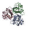
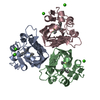
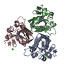
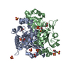
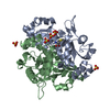
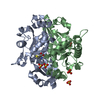
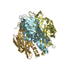
- PDB-2std: SCYTALONE DEHYDRATASE COMPLEXED WITH TIGHT-BINDING INHIBITOR CARP... -
+ Open data

- Basic information
| Entry | Database: PDB / ID: 2std | ||||||
|---|---|---|---|---|---|---|---|
| Title | SCYTALONE DEHYDRATASE COMPLEXED WITH TIGHT-BINDING INHIBITOR CARPROPAMID | ||||||
 Components Components | SCYTALONE DEHYDRATASE | ||||||
 Keywords Keywords | LYASE / MELANINE BIOSYNTHESIS | ||||||
| Function / homology |  Function and homology information Function and homology informationscytalone dehydratase / scytalone dehydratase activity / melanin biosynthetic process / endosome / metal ion binding Similarity search - Function | ||||||
| Biological species |  Magnaporthe grisea (fungus) Magnaporthe grisea (fungus) | ||||||
| Method |  X-RAY DIFFRACTION / Resolution: 2.1 Å X-RAY DIFFRACTION / Resolution: 2.1 Å | ||||||
 Authors Authors | Nakasako, M. / Motoyama, T. / Kurahashi, Y. / Yamaguchi, I. | ||||||
 Citation Citation | Journal: Biochemistry / Year: 1998 Title: Cryogenic X-ray crystal structure analysis for the complex of scytalone dehydratase of a rice blast fungus and its tight-binding inhibitor, carpropamid: the structural basis of tight-binding inhibition. Authors: Nakasako, M. / Motoyama, T. / Kurahashi, Y. / Yamaguchi, I. | ||||||
| History |
|
- Structure visualization
| Structure viewer | Molecule: MolmilJmol/JSmol |
|---|
- Downloads & links
-Download
| PDBx/mmCIF format | 2std.cif.gz | 51.1 KB | Display | PDBx/mmCIF format |
|---|---|---|---|---|
| PDB format | pdb2std.ent.gz | 36.3 KB | Display | PDB format |
| PDBx/mmJSON format | 2std.json.gz | Tree view | PDBx/mmJSON format | |
| Others |  Other downloads Other downloads |
-Validation report
| Arichive directory | https://data.pdbj.org/pub/pdb/validation_reports/st/2stdftp://data.pdbj.org/pub/pdb/validation_reports/st/2std | HTTPS FTP |
|---|
-Related structure data
| Related structure data |  1stdS S: Starting model for refinement |
|---|---|
| Similar structure data |


-Links
 PDBj
PDBj
- Assembly
| Deposited unit | 
| ||||||||
|---|---|---|---|---|---|---|---|---|---|
| 1 | 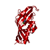
| ||||||||
| Unit cell |
|
-Components
| #1: Protein | Mass: 20280.902 Da / Num. of mol.: 1 Source method: isolated from a genetically manipulated source Source: (gene. exp.) Magnaporthe grisea (fungus) / Production host:  |
|---|---|
| #2: Chemical | ChemComp-SO4 /   Mass: 96.063 Da / Num. of mol.: 1 / Source method: obtained synthetically / Formula: SO4 Mass: 96.063 Da / Num. of mol.: 1 / Source method: obtained synthetically / Formula: SO4 |
| #3: Chemical | ChemComp-CRP / ((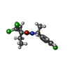  Mass: 334.669 Da / Num. of mol.: 1 / Source method: obtained synthetically / Formula: C15H18Cl3NO Mass: 334.669 Da / Num. of mol.: 1 / Source method: obtained synthetically / Formula: C15H18Cl3NO |
| #4: Water | ChemComp-HOH /  Mass: 18.015 Da / Num. of mol.: 174 / Source method: isolated from a natural source / Formula: H2O Mass: 18.015 Da / Num. of mol.: 174 / Source method: isolated from a natural source / Formula: H2O |
-Experimental details
-Experiment
| Experiment | Method: X-RAY DIFFRACTION / Number of used crystals: 1 |
|---|
- Sample preparation
| Crystal | Density Matthews: 3.5 Å3/Da / Density % sol: 65 % Description: THE CRYSTAL IS NEARLY ISOMORPHOUS WITH THAT OF 1STD | |||||||||||||||||||||||||
|---|---|---|---|---|---|---|---|---|---|---|---|---|---|---|---|---|---|---|---|---|---|---|---|---|---|---|
| Crystal grow | pH: 5.2 / Details: pH 5.2 | |||||||||||||||||||||||||
| Crystal grow | *PLUS Temperature: 20 ℃ / Method: vapor diffusion, hanging drop | |||||||||||||||||||||||||
| Components of the solutions | *PLUS
|
-Data collection
| Diffraction | Mean temperature: 100 K |
|---|---|
| Diffraction source | Source: ROTATING ANODE / Type: RIGAKU ULTRAX 18 / Wavelength: 1.5418 |
| Detector | Type: RIGAKU RAXIS IV / Detector: IMAGE PLATE / Date: Oct 18, 1996 / Details: DOUBLE MIRROR FOCUSING |
| Radiation | Monochromator: NI FILTER / Monochromatic (M) / Laue (L): M / Scattering type: x-ray |
| Radiation wavelength | Wavelength: 1.5418 Å / Relative weight: 1 |
| Reflection | Resolution: 2.1→71.1 Å / Num. obs: 12014 / % possible obs: 85.6 % / Observed criterion σ(I): 1 / Redundancy: 4.18 % / Rmerge(I) obs: 0.073 / Net I/σ(I): 14.5 |
| Reflection shell | Resolution: 2.1→2.25 Å / Redundancy: 2.51 % / Rmerge(I) obs: 0.17 / Mean I/σ(I) obs: 3 / % possible all: 74.6 |
| Reflection | *PLUS Num. measured all: 50287 |
| Reflection shell | *PLUS Lowest resolution: 2.2 Å / % possible obs: 74.6 % |
- Processing
| Software |
| ||||||||||||||||||||||||||||||||||||||||||||||||||||||||||||
|---|---|---|---|---|---|---|---|---|---|---|---|---|---|---|---|---|---|---|---|---|---|---|---|---|---|---|---|---|---|---|---|---|---|---|---|---|---|---|---|---|---|---|---|---|---|---|---|---|---|---|---|---|---|---|---|---|---|---|---|---|---|
| Refinement | Starting model: 1STD Resolution: 2.1→8 Å / Data cutoff high absF: 1000 / Data cutoff low absF: 1 / Isotropic thermal model: GAUSS / σ(F): 2
| ||||||||||||||||||||||||||||||||||||||||||||||||||||||||||||
| Displacement parameters | Biso mean: 28.5 Å2 | ||||||||||||||||||||||||||||||||||||||||||||||||||||||||||||
| Refine analyze | Luzzati coordinate error obs: 0.25 Å | ||||||||||||||||||||||||||||||||||||||||||||||||||||||||||||
| Refinement step | Cycle: LAST / Resolution: 2.1→8 Å
| ||||||||||||||||||||||||||||||||||||||||||||||||||||||||||||
| Refine LS restraints |
| ||||||||||||||||||||||||||||||||||||||||||||||||||||||||||||
| LS refinement shell | Resolution: 2.1→2.19 Å / Total num. of bins used: 8
| ||||||||||||||||||||||||||||||||||||||||||||||||||||||||||||
| Xplor file |
| ||||||||||||||||||||||||||||||||||||||||||||||||||||||||||||
| Software | *PLUS Name: X-PLOR / Classification: refinement | ||||||||||||||||||||||||||||||||||||||||||||||||||||||||||||
| Refinement | *PLUS | ||||||||||||||||||||||||||||||||||||||||||||||||||||||||||||
| Solvent computation | *PLUS | ||||||||||||||||||||||||||||||||||||||||||||||||||||||||||||
| Displacement parameters | *PLUS | ||||||||||||||||||||||||||||||||||||||||||||||||||||||||||||
| Refine LS restraints | *PLUS
| ||||||||||||||||||||||||||||||||||||||||||||||||||||||||||||
| LS refinement shell | *PLUS Rfactor obs: 0.252 |
